Epididymal Cystic Lymphangioma Presenting as Scrotal Swelling in a Post Surgery Case of Carcinoma Rectum- A Case Report
Mohammad Haroon1, Yashmin Nisha2, Kashif Iqubal3
1 Senior Resident, Department of Radiology, Govind Ballabh Pant Institute of Postgraduate Medical Education, New Delhi, India.
2 Junior Resident, Department of Radiology, Rajiv Gandhi Cancer Institute and Research Centre, New Delhi, India.
3 Senior Resident, Department of Paediatrics, Aruna Asaf Ali Hospital, New Delhi, India.
NAME, ADDRESS, E-MAIL ID OF THE CORRESPONDING AUTHOR: Dr. Mohammad Haroon, NP 145B, Pitampura, Near Gopal Mandir, New Delhi-110034, India.
E-mail: haroon.radiology@gmail.com
Cystic lymphangiomas are usually congenital malformations of draining lymphatic channels with most common sites including neck, axilla, mediastinum and retroperitoneum. Occurrence of lymphangiomas in scrotum or inguinal region is a rare entity and epididymal origin of these lesions is even more infrequent. We herein report a case of epididymal lymphangioma detected on USG, which developed after surgical abdominal lymph nodal dissection in an adult patient of carcinoma rectum presenting as painless scrotal swelling.
Epididymis, Lymphatic, Rectum, Scrotum
Case Report
A 24-year-old male presented to the institute in GI Oncosurgery Department as a diagnosed case of metastatic adenocarcinoma of rectum. The cross sectional imaging done in the radiology department also showed evidence of polypoidal infiltrative rectal mass with perirectal, bilateral iliac and paraaortic lymphadenopathy and bony lesions [Table/Fig-1]. Patient underwent abdominoperineal resection with abdominal lymph node dissection after receiving neoadjuvant chemotherapy and radiotherapy. Thereafter, patient received multiple cycles of FOLFOX-4 based chemotherapy and was on regular follow up for one and a half years, when he presented with complain of painless right scrotal swelling. USG examination revealed multicystic septated lesion involving head and body of right epididymis measuring 3.0 x 1.6 x 3.6 cm. On colour Doppler imaging there was no internal vascularity [Table/Fig-2a,b]. Mild ipsilateral hydrocele was also present. Bilateral testis were unremarkable. Imaging differentials of cystic lymphangioma, loculated hydrocele, epididymal cysts and spermatocele were considered and patient underwent excisional biopsy. Histopathological evaluation was suggestive of epididymal lymphangioma as evidenced by microscopic findings of network of multiple irregular dilated cystic spaces lined by single layer of endothelium with stromal infiltration by lymphocytes. Postoperative period was uneventful and the patient was discharged in stable condition. Follow up with USG imaging after three months showed no evidence of recurrence.
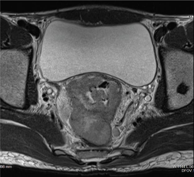
High resolution T2W axial image showing infiltrative malignant rectal mass (arrow) with perirectal lymphadenopathy.
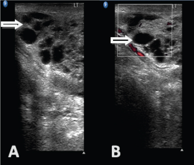
A) Grey scale image showing multicystic septated lesion (arrow) involving head and body of right epididymis; and B) Colour Doppler image showing no internal vascularity (arrow).
Discussion
Lymphangiomas are benign lymphatic malformations mostly presenting in paediatric population and consist of approximately 26% of all benign vascular neoplasms in children [1]. Occurrence of these lesions is infrequent in adults. The most common sites for children as well as adults are head/neck, axilla, mediastinal and retroperitoneal regions. Scrotum and inguinal canal are uncommon sites for development of lymphangiomas as only about 20 cases have been reported in literature [2]. Epididymal lymphangiomas are even more infrequently encountered, to the best of our knowledge, only six cases have been reported so far with the first case reported by Thompson in 1936 [3].
Epididymal lymphangioma can occur either as congenital true primary neoplasm or secondary to infection, trauma or post-surgical lymphatic blockage. Out of previously reported 6 cases of epididymal lymphangioma one case was thought to develop secondary neoplasma to herniorrhaphy [4] and rest of the five cases were diagnosed as true primary neoplasms [3,5-8].
In 1976, Whimster first elaborated lymphangiomas, he postulated these lesions to be congenital abnormalities due to anomalous development of the lymphatic channels, originating from sequestered lymphatic tissue which does not develop a normal communication with lymphatic system and thus, present as mass later on [9].
The imaging differentials of cystic epididymal mass lesions mainly consist of epdidymal cysts, spermatoceles and loculated hydrocele. Epididymal cysts and spermatoceles are usually unilocular lesions while lymphangiomas are multicystic septated lesions. However, imaging may be suggestive but definitive diagnosis can only be made by histopathological examination. These lesions frequently show recurrence due to incomplete surgical excision in 25-30% of the cases [10,11].
We herein present a case of right epididymal lymphangioma in a patient of metastatic carcinoma rectum on palliative chemotherapy. Around two years ago, the patient underwent abdominoperineal resection with iliac and paraaortic lymphnode dissection. This patient presented to our institute with painless right scrotal swelling, underwent USG and histopathological evaluation which was finally diagnosed as epididymal lymphangioma possibly resulting as sequelae to previous surgery two years back. Follow up USG examination didn’t show any recurrence. To the best of our knowledge, only one such case has been described in literature, where lymphangioma of the epididymis was thought to develop secondarily to previous herniorrhaphy surgery [4].
Our case report is unique in several aspects. First of all lymphangiomas in general are rare in adult patients, inguinoscrotal lymphangiomas are even rarer. A case of adult lymphangiomas, arising from the epidydimis, secondary to surgery is only the second case reported in literature till now, to the best of author’s knowledge.
Conclusion
The purpose of our case report is to familiarize the radiologists with this rare entity. Although extremely rare, differential of cystic lymphangiomas should also be considered in a patient presenting with scrotal swelling with previous history of surgery.
[1]. Jamal YS, Rahman A, Halim A, Moshref SS, Kurdi MO, Sandugji HI, Cystic lymphangioma of spermatic cord: A case report and literature reviewJKAU Med Sci 2009 16:103-11. [Google Scholar]
[2]. Rastogi R, Meena GL, Kumar R, Rastogi V, Cystic lymphangioma scroti: A common tumor at a rare locationSaudi J Kidney Dis Transpl 2010 21:1132-34. [Google Scholar]
[3]. Thompson GJ, Tumors of the spermatic cord, epididymis and testicular tunicsSurg Gynecol Obstet 1936 62:712 [Google Scholar]
[4]. Postius J, Manzano C, Concepcion T, Castro D, Gutierrez P, Banares F, Epididymal lymphangiomaJ Urol 2000 163:550-51. [Google Scholar]
[5]. Kini H, Minal J, Prabhu LG, Suresh PK, Basavaiah SH, Epididymal lymphangioma: An unusual cause for retro testicular mass in an adultMed J DY Patil Univ 2015 8:819-21. [Google Scholar]
[6]. Pai KK, Roy D, Lymphangioma of epididymis: An extremely rare cause of scrotal swellingIndian J Urol 2006 22:275-76. [Google Scholar]
[7]. Enzinger FM, Weiss SW, Tumors of lymph vesselsIn : soft tissue tumors 1988 2nd edMosby CoSt. Louis:614 [Google Scholar]
[8]. Edwards WD, Wold LE, Congenital lymphangiectasis. In : Coulson WF (editor)Surgical pathology 1988 PhiladelphiaLippincott Co:499 [Google Scholar]
[9]. Whimster IW, The pathology of lymphangioma circumscriptumBr J Dermatol 1976 94(5):473-86. [Google Scholar]
[10]. Haroon S, Hasan SH, Lymphangioma circumscriptum in the scrotum: A case reportJ Med Case Rep 2012 6:233 [Google Scholar]
[11]. Olabanji J, Oladele A, Famurewa O, Adejuyigbe O, Ademola S, Retroperitoneal and genital lymphangioma therapeutic challenges in a developing countryLibyan J Med 2009 4:44-45. [Google Scholar]