Cervical cancer is the most common cancer among Indian women between 15 and 44 years of age. Current estimates indicate that every year 13,4420 women are diagnosed with cervical cancer and 72,825 die from the disease [1]. The vast majority of cervical cancer cases are caused by infection with certain subtypes of HPV, a sexually transmitted virus that infects cells and may result in precancerous lesions and invasive cancer [2]. At a given time in the general population about 7.9% of women are estimated to have cervical HPV infection, and HPV 16 or 18 are attributed to 82.5% of invasive cervical cancers [1]. Genomic integration of the viral genome can disrupt several cellular proteins resulting in their upregulation. One of the consequences is the upregulation of the tumour suppressor gene p16INK4A which is a cyclin dependent kinase inhibitor [3]. The protein p16 is integral to Rb (retino-blastoma) mediated counters of the G1-S phase transition of the cell cycle by inactivating cyclin dependent kinases that phosphorylate Rb protein. A reciprocal relation between p16 and Rb has been observed. The E7 protein of the HPV binds and inactivates Rb which leads to the release of E2F, a transcription factor, which in turn can activate the genes required for entry into S-phase of the cell cycle. This results in increased levels of p16 through a negative feedback mechanism. Interference of the viral oncoproteins with cellular proteins involved in cell cycle regulation indicates overexpression of p16 [4].
The protein p16INK4A is overexpressed in cervical intraepithelial neoplasia and invasive cancer and serves as a surrogate marker for the oncogenic activities of HPV in replication-competent cells of cervical epithelia. There are very few reports on p16INK4A in cervical dysplasia and cancer in Indian literature, despite the fact that Indian females represent a major proportion of the affected population [3]. This study was conducted as a prospective study to evaluate the usefulness of p16INK4A as a biomarker in dysplastic and malignant alteration of cervical epithelium.
Materials and Methods
This prospective study of two years duration (from August 2013 to July 2015) was conducted in the Department of Pathology, M. R. Medical College, Kalaburagi, Karnataka, India. Ethical clearance for this study was taken from Institute Ethics Board.
A total of 57 cervical biopsies and hysterectomies were collected out of which 47 cases were of carcinoma cervix and 10 cases were of CINs. Twenty randomly selected cases of normal (non dysplastic) cervical epithelium were studied to know the negative association with p16INK4A (control group).
All hysterectomy or cervical biopsy specimens from patients diagnosed with cervical neoplasms were included in the study. Patients having recurrence of cervical malignancies and undergoing treatment and patients vaccinated against HPV were excluded.
Cervical dysplasia was classified using CIN classification [5]. Cervical carcinoma was classified according to WHO histological classification of invasive carcinomas of uterine cervix [6].
SCC cervix was sub typed into keratinizing and non keratinizing types [7,8]. Broder’s grading was used for grading SCC cervix [9]. Adenocarcinoma cervix was graded as well differentiated, moderately differentiated and poorly differentiated types [10].
IHC
All the cases diagnosed histologically as carcinoma cervix or CIN were subjected to IHC study for p16INK4A expression using super sensitive polymer-Horseradish Peroxidase (HRP) based immunohistochemistry kit of BioGenex (G175-405). Twenty randomly selected cases of hysterectomies done for other gynaecologic causes were used to study the expression of p16INK4A in normal (non dysplastic) cervical epithelium. These were found to have statistically comparable parameters like age and parity, required for appropriate comparison with the cases. Cases of squamous cell carcinoma served as positive control, control tissue for which was provided by BioGenex (FG-540M). P16INK4A Immunostaining was evaluated using two different scoring protocols [3,11].
Scoring criteria 1: Positive (moderate or strong staining in more than 10% of epithelial cells).
Negative (less than 10% of epithelial cells with weak, moderate or strong staining).
Scoring criteria 2: A semi-quantitative IHC score [11,12].
Points were given according to Intensity of staining [11]: 0 = no staining; 1 = weak staining; 2 = moderate staining; 3 = strong staining
Points were given according to proportion of cells stained [11,12]: 0 = no staining; 1 = <1%; 2 = 1-10%; 3 = 11-33%; 4 = 34-66%; 5 = >66%
Score was calculated by adding the points for intensity and proportion of cells stained with p16INK4A immunostain. Maximum score was 8 and Minimum score was 0. Score of 0-2 was taken as low, score 3-5 as moderate while score of 6-8 was taken as overexpression and was used for categorization in various histological types [11,12] after establishing its statistical significance by the above mentioned tests.
Statistical Analysis
The p-value of less than 0.05 was considered statistically significant using SPSS 16.0 software and the descriptive statistics, Chi-square test. The p-value of <0.05 was considered statistically significant.
Results
The p16INK4A expression was absent in normal (non dysplastic) cervical epithelium in all cases and it increased from 25% positive cases in CIN 1(1/4) to 75% (3/4) cases of CIN 3 showing positivity [Table/Fig-1].
Overall pattern of p16INK4A expression in various studies.
| Study | Normal epithelium | CIN 1 | CIN 2 | CIN 3 | Carcinoma cervix |
|---|
| Agoff SN et al., [13] | 11% | 57% | 75% | 91% | 92% |
| (19/33) | (43/76) | (60/80) | (103/113) | (42/46) |
| Murphy N et al., [14] | ND | 100% | 100% | 98% | 100% |
| (38/38) | (33/33) | (45/46) | (10/10) |
| Hu L et al., [15] | ND | 44% | 93% | 100% | ND |
| (20/45) | (43/46) | (51/51) |
| Benevolo M et al., [16] | ND | 31% | 90% | ND | 100% |
| (17/54) | (9/10) | (11/11) |
| Kong CS et al., [17] | ND | 92% | 100% | ND | ND |
| (7/12) | (16/16) |
| Focchi GR et al., [18] | 12% | 91% | 100% | ND | 100% |
| (7/58) | (80/88) | (65/65) | (47/47) |
| Lesnikova Iana et al., [11] | 00 | 72.3% | 91% | 98.3% | 98.5% |
| (0/10) | (180/249) | (212/233) | (178/181) | (131/133) |
| Gupta R et al., [3] | 10% | 50% | 60% | 70% | 95% |
| (2/20) | (10/20) | (12/20) | (14/20) | (19/20) |
| Srivastava S [19] | 00 | 80% | 100% | 100% | 100% |
| (0/15) | (8/10) | (5/5) | (3/3) | (15/15) |
| Kumari K and Vadivelan AA [20] | 00 | 62.5% | 75% | 81.25% | 100% |
| (0/16) | (10/16) | (12/16) | (13/16) | (16/16) |
| Present study | 00 | 25% | 50% | 75% | 100% |
| (0/20) | (1/4) | (1/2) | (3/4) | (47/47) |
ND- not done
On semi-quantitative scoring, over expression (score>5) for p16INK4A was observed in 25% cases of CIN 1, 50% cases of CIN 2 and 75% cases of CIN 3 (p<0.05). All cases of carcinoma cervix were positive for p16INK4A expression [Table/Fig-2], 73.33% of SCC and all cases of adenocarcinoma showed over expression for p16INK4A with score >5.
p16INK4A expression in carcinoma cervix in various studies.
| Study | Cases | Positive | Negative |
|---|
| Volgareva G et al., [21] | 21 (SCC) | 95.23% | 4.77% |
| 5 (Adenocarcinoma) | 100% | 00 |
| Murphy N et al., [22] | 10 (SCC) | 100% | 100% |
| 10 (Adenocarcinoma) | 00 | 00 |
| Lesnikova Iana et al., [11] | 133 | 98.5% | 1.5% |
| Gupta R et al., [3] | 20 | 95% | 5% |
| Tan GC et al., [23] | 72 | 98.6% | 1.4% |
| Srivastava S [19] | 15 | 100% | 00 |
| Kumari K and Vadivelan AA [20] | 16 | 100% | 00 |
| Present study | 45 (SCC) | 100% | 00 |
| 02 (Adenocarcinoma) | 100% | 00 |
About 85% cases of keratinizing SCC and 64% cases of non keratinizing SCC type showed over expression of p16INK4A.; 93.75% case of Grade I, 60.86% cases of Grade II and 66.66% cases of Grade III SCC showed over expression of p16INK4A with score >5. Both cases of adenocarcinoma also showed over expression of p16INK4A.
Expression of p16INK4A in all these entities has been shown in [Table/Fig-3,4,5].
a) p16INK4A negative staining (DAB, 40X); b) Weak p16INK4A staining (DAB, 40X); c) Moderate p16INK4A staining (DAB, 40X); d) Strong p16INK4A staining (DAB, 40X).
*DAB: 3,3’-Diaminobenzidine

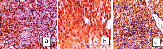
a) Strong p16INK4A staining in Well differentiated SCC- keratinzing type (DAB, 40X) b) Strong p16INK4A staining in moderately differentiated SCC (DAB, 40X) c) Strong p16INK4A staining in poorly differentiated SCC (DAB, 10X).
*DAB: 3,3’-Diaminobenzidine
a) Moderate p16INK4A staining in well differentiated adenocarcinoma cervix (DAB,10X) b) Moderate p16INK4A staining in poorly differentiated adenocarcinoma cervix (DAB, 40X).
*DAB: 3,3’-Diaminobenzidine

Discussion
On analysing the overall pattern among all the cases studied for p16INK4A expression in this study, all the cases of normal (non-dysplastic) cervical epithelium showed a low expression score of range taken as 0-2, 25% cases of CIN 1, 50% cases of CIN 2, 75% cases of CIN 3 and 74.46% cases of carcinoma cervix showed over expression of p16INK4A [Table/Fig-3]. The p16INK4A expression was also assessed in 20 randomly selected cases of normal (non dysplastic) cervical epithelium and 100% of cases showed negative expression. The findings are similar to the studies by Lesnikova Iana et al., Srivastava S and Tan GC et al., [11,19,23].
In our study, p16INK4A positivity was noted in 25% cases of CIN1 (1/4), 50% cases of CIN 2 (1/2) and 75% cases of CIN3 (3/4). These findings are in accordance with the similar studies by Gupta R et al., Lesnikova Iana et al., Srivastava S et al., and Kumari K and Vadivelan AA [3,11,19,20]. Overall, 50% (5/10) of the cases of CIN showed positive expression for p16INK4A which was similar to the study by Gupta R et al., [3].
On semi-quantitative scoring, over expression of p16INK4A increased as the cervical dysplasia progressed from CIN1 (25%) to CIN 3 (75%). The findings are in accordance with the study by Lesnikova Iana et al., who also found progressive over expression of p16INK4A from CIN1 (43.3%) to CIN 3 (94%) [11].
Out of 47 cases of carcinoma cervix, 45 were squamous cell carcinoma and two were adenocarcinoma. All the cases showed positive expression for p16INK4A which is similar to the studies by Lesnikova Iana et al., (98.5%), Srivastava S (100%) Kumari K and Vadivelan AA. (100%) Volgareva G et al., (95% in SCC and 100% in adenocarcinoma), and Murphy N et al., (100%) [Table/Fig-2] [11,19-22].
In this study, 74.46% cases of carcinoma cervix showed over expression of p16INK4A on semi quantitative scoring while 25.53% showed moderate score. The findings are similar to the study by Lesnikova Iana et al., [11].
On tabulating the overall expression pattern for p16INK4A, normal (non-dysplastic) cervical epithelium showed negative expression in all the cases. As the dysplasia increased from CIN 1 to CIN 3, the p16INK4A positivity also increased from 25% to 75%. All the cases of carcinoma cervix showed positive expression. The These findings are similar to other such studies by Gupta R et al., Lesnikova Iana et al., Murphy N et al., Benevolo M et al., Focci GR et al., Srivastava S and Kumari K and Vadivelan AA [Table/Fig-1] [3,11,14,16,18-20].
Limitation
Paucity of samples was the reason for the small sample size in this study but the findings suggest a positive outcome and this could be taken as a prospective study for further research using a bigger sample size over a longer duration.
Conclusion
In this study, two types of scoring patterns were used and compared. One was a simple positive versus negative score. Other was a detailed but still easy to apply method in which semi quantitative immunohistological scoring system of p16INK4A was done. The latter gave a more detailed picture of the spread of expression scores among various parameters associated with cervical carcinoma and CIN by showing the expression patterns among various histological grades of cervical cancer. Significant over expression of p16INK4A was observed in carcinoma cervix and with increasing severity of cervical dysplasia, the p16INK4A expression increased progressively. The important parameters significantly associated with carcinoma cervix and cervical dysplasia like parity and age at presentation showed significantly positive expression for p16INK4A and gave high expression scores indicating the association HPV with majority of cases of carcinoma cervix in North Karnataka region. This is a prospective study which implicates the efficacy of p16INK4A as a marker for screening cervical malignancies especially in developing countries [15] and possibly as a surrogate marker for HPV infection and emphasizes towards the use of vaccination against HPV infection.
ND- not done