Fracture Resistance of Endodontically Treated Teeth Restored with Biodentine, Resin Modified GIC and Hybrid Composite Resin as a Core Material
Dayalan Subash1, Krishnamma Shoba2, Shibu Aman3, Srinivasan Kumar Indu Bharkavi4, Vijayan Nimmi5, Radhakrishnan Abhilash6
1 Senior Resident, Department of Conservative Dentistry and Endodontics, Government Dental College, Kottayam, Kerala, India.
2 Head, Department of Conservative Dentistry and Endodontics, Government Dental College, Kottayam, Kerala, India.
3 Assistant Professor, Department of Conservative Dentistry and Endodontics, Government Dental College, Kottayam, Kerala, India.
4 Senior Lecturer, Department of Oral Pathology and Microbiology, Sathyabama University Dental College and Hospital, Chennai, Tamil Nadu, India.
5 Junior Resident, Department of Conservative Dentistry and Endodontics, Government Dental College, Kottayam, Kerala, India.
6 Junior Resident, Department of Conservative Dentistry and Endodontics, Government Dental College, Kottayam, Kerala, India.
NAME, ADDRESS, E-MAIL ID OF THE CORRESPONDING AUTHOR: Dr. Dayalan Subash, NO.53, Bharathiyar Street, Tiruttani-631209, Tamil Nadu, India.
E-mail: dr.subash06@yahoo.com
Introduction
The restoration of a severely damaged tooth usually needs a post and core as a part of treatment procedure to provide a corono - radicular stabilization. Biodentine is a class of dental material which possess high mechanical properties with excellent biocompatibility and bioactive behaviour. The sealing ability coupled with optimum physical properties could make Biodentine an excellent option as a core material.
Aim
The aim of the study was to determine the fracture resistance of Biodentine as a core material in comparison with resin modified glass ionomer and composite resin.
Materials and Methods
Freshly extracted 30 human permanent maxillary central incisors were selected. After endodontic treatment followed by post space preparation and luting of Glass fibre post (Reforpost, Angelus), the samples were divided in to three groups based on the type of core material. The core build-up used in Group I was Biodentine (Septodont, France), Group II was Resin-Modified Glass Ionomer Cement (GC, Japan) and Group III was Hybrid Composite Resin (TeEconom plus, Ivoclar vivadent). The specimens were subjected to fracture toughness using Universal testing machine (1474, Zwick/Roell, Germany) and results were compared using One-way analysis of variance with Tukey’s Post hoc test.
Results
The results showed that there was significant difference between groups in terms of fracture load. Also, composite resin exhibited highest mean fracture load (1039.9 N), whereas teeth restored with Biodentine demonstrated the lowest mean fracture load (176.66 N). Resin modified glass ionomer exhibited intermediate fracture load (612.07 N). The primary mode of failure in Group I and Group II was favourable (100%) while unfavourable fracture was seen in Group III (30%).
Conclusion
Biodentine, does not satisfy the requirements to be used as an ideal core material. The uses of RMGIC’s as a core build-up material should be limited to non-stress bearing areas. Composite resin is still the best core build-up material owing to its high fracture resistance and bonding to tooth.
Compressive strength, Corono-radicular stabilization, Post and core
Introduction
A multitude of injuries often result in considerable coronal hard tissue defects, requiring post and core as a preprosthetic treatment, providing retention and support for the extracoronal prosthesis restoring the lost function and aesthetics [1–3]. A variety of core materials are being used these days out of which cast gold, amalgam, resin-based composite and glass ionomer cement are the most popular. Amalgam requires prolonged setting time, preventing crown preparation in the same visit [4]. A cast gold post and core, however, it is an indirect procedure requiring two visits.
Resin-modified glass ionomer cements set rapidly, after chemical- or light initiation, allowing for an immediate finishing of the restoration with better mechanical properties [5–7]. On the contrary, their greater degree of shrinkage upon polymerization compared to conventional glass ionomers [5], lower rigidity compared to that of composites [8], and strength lower than that of the tooth structure, are major drawbacks [2,9]. In addition, resin-modified glass ionomers also lacks translucency [7].
Resin composites, are commonly used as a core build –up material because of its high mechanical properties and bonding ability to tooth. On the contrary, these materials display technique sensitivity and are time consuming [5,10]. Moreover, inadequate Degree of Conversion (DC), inherent polymerization shrinkage, resulting in breakdown at the interface and consequent gap formation causing microleakage leading to failure [11]. The huge potential for water- uptake and the high coefficient of thermal expansion are other shortcomings of these materials [2].
In 2009, synthetic tricalcium silicate cement (Biodentine, Septodont, SaintMaur-des-Fossées, France) became commercially available. The use of bioactive materials as core materials opens new prospects in restorative dentistry as they bond to the tooth naturally and can heal coronal as well as furcal perforations when present. Biodentine has been developed and produced with the aim of bringing together the high biocompatibility and bioactivity of calcium silicates, with enhanced properties such as quick setting time (in comparison with MTA) and high strength properties not usually associated with other tricalcium cements [12]. Raskin A et al., found that the marginal sealing was equivalent to that of the resin-modiifed glass ionomer cement (Fuji II LC) restorations [13].
The sealing ability coupled with optimum physical properties could make Biodentine an excellent option as a core material. Therefore, it becomes worthwhile to study the fracture resistance of Biodentine in comparison with proven core materials like resin-modified glass ionomer cement and composite resin.
Materials and Methods
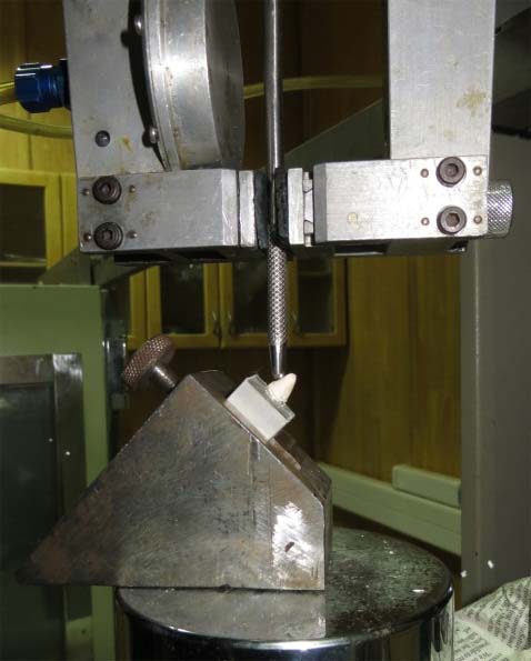
This is an in vitro study conducted in the Department of Conservative dentistry and Endodontics, Government Dental College, Kottayam and Rubber Research Institute of India, Kottayam, Kerala, India. The following core materials were used: Biodentine (Septodont, France), Glass ionomer light curable universal restorative Type II (GC, Japan), Te- Econom plus (Ivoclarvivadent, Liechtenstein). A total of 30 recently extracted single-rooted human maxillary central incisors were used. All samples were prepared by a single operator. Endodontic treatment was done in all the 30 samples. Tooth samples were mounted in acrylic resin mould and then crowns decoronated leaving 3 mm of crown structure above cementenamel junction. After post space preparation glass fibre post (Reforpost (Angelus, Brazil)) of 1.1 mm thickness was luted with Resin cement {Multilink Speed (Ivoclar vivadent, Liechtenstein)}. Samples were divided into three groups with 10 samples in each group. Core materials were mixed according to their manufacturer’s instructions and packed inside the strip crown no.4.The core build-up used in Group I was Biodentine, Group II was resin-modified Glass ionomer cement and Group III was hybrid composite resin. Each specimen was held in place for testing in a special jig with its long axis inclined facially, at an angle of 1350 and subjected to a load on a universal testing machine at a crosshead speed of 0.5 mm/minute until failure occurred [Table/Fig-1].
Mounted specimen at an angle of 1300
The reading was noted to determine the maximum force at failure in Newton (N).
Statistical Analysis
The results obtained were tabulated and subjected to One-way ANOVA and Tukey’s post-hoc using Statistical Package for Social Sciences (SPSS, V 16.0) package with significance value (p) kept at <0.05.
Results
The mean peak failure load for Group I, Group II and Group III along with Standard deviation values was 176.66±5.75 N, 612.07±8.00 N, 1039.90±15.94 N respectively. The results showed that Group I samples restored with Biodentine fractured at the lowest load (176.66±5.75N) applied, while Group III samples restored with composite fractured at the highest load (1039.90±15.94N) applied. Group II exhibited intermediate values between the two groups. To compare the mean failure loads difference among the groups, one-way ANOVA [Table/Fig-2] and Tukey’s post-hoc test at 95% level of significance was employed [Table/Fig-3]. Intergroup comparison of these results shows that, there is statistically significant (p < 0.001) difference among the groups. In term of fracture mode, majority of the samples were fractured near the cementoenamel junction in the oblique direction.
Comparison of mean failure loads difference among groups using one way ANOVA.
| Groups | N | Mean failure load (in Newton) | Std. Deviation | F value | p-value |
|---|
| Group I | 10 | 176.66 | 5.75 | 0.0159 | <0.001 |
| Group II | 10 | 612.07 | 8.00 |
| Group III | 10 | 1039.90 | 15.94 |
Inter Group comparison using Tukey’s Post-Hoc.
| Group | Group | Mean Difference (N) | Std. Error | Sig. (p-value). | 95% Confidence interval |
|---|
| Lower bound | Upper bound |
|---|
| I | II | -435.41300* | 4.84083 | <.001 | -447.4154 | -423.4106 |
| III | -863.24200* | 4.84083 | <.001 | -875.2444 | -851.2396 |
| II | I | 435.41300* | 4.84083 | <.001 | 423.4106 | 447.4154 |
| III | -427.82900* | 4.84083 | <.001 | -439.8314 | -415.8266 |
| III | I | 863.24200* | 4.84083 | <.001 | 851.2396 | 875.2444 |
| II | 427.82900* | 4.84083 | <.001 | 415.8266 | 439.8314 |
1p-value <0.001 implies difference between are statistically significant
2The table shows that the comparison between Group I, Group II and Group III has a p-value of < 0.001, which is <0.05. So, the mean failure loads between groups were statistically significant.
Discussion
The fracture resistance of root post-core assembly is of paramount importance for long term stability of the restoration. Stress was generated within the body opposing the external force to prevent fracture. When this force exceeds the internal stress it results in fracture [14,15]. Therefore, when stress exceeds the cohesive strength of the object; the object breaks [16]. In this study, Biodentine group showed significantly lower fracture resistance than resin-modified GIC and composite. It seems that the compressive forces applied on specimens restored with Biodentine have created stresses within the specimens that exceeded the cohesive strength of these specimens and lead to earlier failure at lower load values compared to other groups.
Compressive strength is considered critical because it is what resists masticatory and parafunctional forces [17]. Biodentine is also called as ‘dentin substitute’. Manufacturer claims, it possess compressive strength of 300 MPa (dentin – 297 MPa), flexural strength of 24 MPa, elastic modulus of 22 GPa (dentin – 18.5 GPa), Vickers microhardness number of 60.9 whereas dentin is 60 [18].
Franquin J-C et al., claimed that Biodentine have compressive strength value of 316.3 MPa after 28 days [19]. However, the results expressed in our study by Biodentine (176.66 N) were in contradiction with these findings. The material exhibiting a compressive strength 300 MPa should have a higher fracture resistance. while resin modified glass ionomer with compressive strength of 200 MPa [20] showed much higher fracture resistance than Biodentine. Jang Y-E et al., studied the physical properties of Biodentine and affirms that the compressive strength to be 61.35±5.09 N which is much inferior to what Franquin J-C et al., claimed in his study [19]. Our study also found inferior value but greater than the value obtained by Jang Y-E et al., [21]. Study conducted by Lucas CD et al., also exhibited inferior mechanical properties for Biodentine [22].
Group II teeth restored with resin-modified glass ionomer cement (612.07 N) had better fracture resistance value than Biodentine, but significantly lower than composite group. These findings were in concord with various studies [17,23–26]. Further, mechanical properties such as in vitro fatigue resistance and compressive strength of glass-ionomer cements were found to be inadequate for use in stress- bearing areas and were considered inappropriate for dowel and core restorations [27]. Martinez- Insua A et al., also obtained similar results and stated that resin-modified glass ionomers should be limited to non–stress-bearing areas [14].
In Group III, teeth restored with hybrid composite resin (1039.9N) had better fracture resistance than Biodentine and resin-modified glass ionomer cement. The use of fibre posts in combination with direct composite resin core materials for endodontically treated mutilated teeth resulted in better resistance form as found by clinical evaluation by Mohan SM et al., [28]. This was in agreement with other clinical studies [29–33]. The introduction of glass fibre posts and composite resin has brought a new concept of “Endoesthetics” into picture. Moreover, glass fibre post is translucent and also creates a monoblock effect, bonding every component directly or indirectly thus, reinforcing the intra-radicular tooth structure with excellent transverse strength [34]. The Monoblock effect has been proven to reduce catastrophic failure of endodontically treated teeth.
The primary mode of failure (90%) for all groups was favourable (fracture line not extending below CEJ) in general. Individual analysis showed unfavourable failures (fracture line extending below CEJ) about 30% in Group III. These findings are in harmony with Akkayan B and Gulmez T study, they too had 40% catastrophic failure in glass fibre post with composite core group [35].
Our study also showed similar results. It needs to be noted that catastrophic (unfavourable) failures occurred when the loading forces were greater than normal physiological masticatory force exerted on maxillary central incisors. The average masticatory forces in the oral cavity were reported in the range of 146.17 N [36], 193 N [37] and 235.9 N [38]. In the present study, unfavourable fractures that found in Group III namely composite cores were in the range of 1039.9 N, which rarely occur in clinical situation. Therefore, results obtained for the unfavourable fractures for composite core should not be interpreted as an adverse effect.
Within the limitations of the study we conclude that Biodentine does not satisfy the requirements to be used as an ideal core material. The uses of resin-modified glass ionomer cement as a core build-up material should be limited to non-stress bearing areas. Composite resin is still the best core build-up material owing to its high fracture resistance and bonding to tooth. Though, Biodentine is inferior when used as core-build up material, it may still find relevance when used as double seal restoration or perforation repair material. Research may be directed to other fields like biocompatibility and sealing ability where Biodentine may perform much better.
Conclusion
Bioactive materials can enhance healing, when placed near biological tissues. We found that it is not an able substitute for core build-up materials in terms of physical properties. Advancement in biomaterials needs to be directed towards developing new materials with improved physical properties.
1p-value <0.001 implies difference between are statistically significant
2The table shows that the comparison between Group I, Group II and Group III has a p-value of < 0.001, which is <0.05. So, the mean failure loads between groups were statistically significant.
[1]. Shillingburg HT, Hobo S, Whitsett LD, Jacobi R, Brackett S, Fundamentals of fixed Prosthodontics 1997 3rd edIllinoisQuint Publishing Co [Google Scholar]
[2]. Walmsley A, Walsh T, Burke F, Shortall A, Lumley P, Hayes-Hall R, Restorative Dentistry 2002 ChicagoChurchill Livingstone [Google Scholar]
[3]. Piwowarczyk A, Ottl P, Lauer H, Büchler A, Laboratory strength of glass ionomer cement, compomers, and resin compositesJ Prosthodont 2002 11(2):86-91. [Google Scholar]
[4]. Sahmali SM, Saygili G, Compressive shear strength of core materials and restoring techniquesInt J Pedod Restor Dent 2000 20(3):277-83. [Google Scholar]
[5]. Anusavice KJ, Phillips’ Science of Dental Materials 2003 11th edMissouriElsevier Health Sciences [Google Scholar]
[6]. Craig RG, Powers JM, Restorative Dental Materials 2002 11th edMissouriMosby Inc [Google Scholar]
[7]. Wilson AD, Resin-Modified Glass-lonomer CementsInt J Prosthodont 1990 3(5):425-30. [Google Scholar]
[8]. Braem MJ, Lambrechts P, Gladys S, Vanherle G, In vitro fatigue behavior of restorative composites and glass ionomersDent Mater 1995 11(2):137-41. [Google Scholar]
[9]. Christensen GJ, Building up tooth preparations for full crowns-2000J Am Dent Assoc 2000 131(4):505-06. [Google Scholar]
[10]. Manhart J, Kunzelmann KH, Chen HY, Hickel R, Mechanical properties and wear behavior of light-cured packable composite resinsDent Mater 2000 16(1):33-40. [Google Scholar]
[11]. Tarle Z, Meniga A, Knezevic A, Sutalo J, Ristic M, Pichler G, Composite conversion and temperature rise using a conventional, plasma arc, and an experimental blue LED curing unitJ Oral Rehabil 2002 29(7):662-67. [Google Scholar]
[12]. Grech L, Mallia B, Camilleri J, Characterization of set intermediate restorative material, biodentine, bioaggregate and a prototype calcium silicate cement for use as root-end filling materialsInt Endod J 2013 46(7):632-41. [Google Scholar]
[13]. Raskin A, Eschrich G, Dejou J, About I, In vitro microleakage of Biodentine as a dentin substitute compared to Fuji II LC in cervical lining restorationsJ Adhes Dent 2012 14(6):535-42. [Google Scholar]
[14]. Martinez-Insua A, Da Silva L, Rilo B, Santana U, Comparison of the fracture resistances of pulpless teeth restored with a cast post and core or carbon-fiber post with a composite coreJ prosthtic Dent 1998 80(5):527-32. [Google Scholar]
[15]. Maccari PCA, Conceição EN, Nunes MF, Fracture resistance of endodontically treated teeth restored with three different prefabricated esthetic postsJ Esthet Restor Dent 2003 15(1):25-31. [Google Scholar]
[16]. Ferracane JL, Materials in dentistry-principles and applications 2001 2nd edPennsylvaniaLippincott Williams & Wilkins [Google Scholar]
[17]. Bayindir F, Yilmaz CB, Comparison of diametral tensile, flexural, and compressive strengths of five core build-up materialsAtatürk Univ J Fac Dent 2007 17(1):18-23. [Google Scholar]
[18]. SeptodontBiodentine scientific file 2010 [Google Scholar]
[19]. Franquin J, Marie O, Bottero M, Koubi G, About I, Physical properties of a new Ca3SiO5-based dentin substitutePostersessions presented at: IADR Congress, 14-17 July 2010 Barcelona [Google Scholar]
[20]. Aratani M, Pereira AC, Correr-Sobrinho L, Sinhoreti MAC, Consani S, Compressive strength of resin-modified glass ionomer restorative material : effect of P / L ratio and storage timeJ Appl oral Sci 2005 13(4):356-59. [Google Scholar]
[21]. Jang Y-E, Lee B-N, Koh J-T, Park Y-J, Joo N-E, Chang H-S, Cytotoxicity and physical properties of tricalcium silicate-based endodontic materialsRestor Dent Endod 2014 39(2):89-94. [Google Scholar]
[22]. Lucas CD, Viapiana R, Bosso-Martelo R, Guerreiro-Tanomaru JM, Camilleri J, Tanomaru-Filho M, Physicochemical properties and dentin bond strength of a tricalcium silicate-based retrograde materialBrazilian Dental Journal 2017 28(1):51-56. [Google Scholar]
[23]. Bonilla ED, Mardirossian G, Caputo AA, Fracture toughness of various core build-up materialsJ Prosthodont 2000 9(1):14-18. [Google Scholar]
[24]. Jayanthi N, Vinod V, Comparative evaluation of compressive strength and flexural strength of conventional core materials with nanohybrid composite resin core material an in vitro studyJ Indian Prosthodont Soc 2013 13(3):281-89. [Google Scholar]
[25]. Saygili G, Sahmali SM, Comparative study of the physical properties of core materialsInt J Periodontics Restor Dent 2002 22(4):355-63. [Google Scholar]
[26]. Lee S, Lee S, Ph D, Yang J, Han J, Lee J, Fracture toughness of various core materialsJ Korean Acad Prosthodont 2001 39(6):682-97. [Google Scholar]
[27]. Gladys S, Van Meerbeek B, Braem M, Lambrechts P, Vanherle G, Comparative physico-mechanical characterization of new hybrid restorative materials with conventional glass-ionomer and resin composite restorative materialsJ Dent Res 1997 76(4):883-94. [Google Scholar]
[28]. Murali Mohan S, Mahesh Gowda E, Shashidhar MP, Clinical evaluation of the fiber post and direct composite resin restoration for fixed single crowns on endodontically treated teethMed J Armed Forces India 2015 71(3):259-64. [Google Scholar]
[29]. Zicari F, Lesaffre E, An up to 3-Year controlled clinical trial comparing the outcome of glass fiber posts and composite cores with gold alloy–based posts and cores for the restoration of endodontically treated teethInt J Prosthodont 2011 24(4):09-11. [Google Scholar]
[30]. Shashikala K, Sonali S, Clinical and radiological evaluation of cast metal and quartz fibre posts in endodontically restored teethEndodontology 2010 :37-46. [Google Scholar]
[31]. Butz F, Dent M, Heydecke G, Survival rate and fracture strength of endodontically treated maxillary incisors with moderateInt J Prosthodont 2001 14(1):58-64. [Google Scholar]
[32]. Piovesan EM, Demarco F, Survival rates of endodontically treated teeth restored with fiber-reinforced custom posts and cores: A 97-month studyInt J Prosthodont 2007 20(6):633-40. [Google Scholar]
[33]. Cagidiaco MC, Radovic I, Simonetti M, Clinical performance of fiber post restorations in endodontically treated teeth :2-Year ResultsInt J Prosthodont 2007 20(3):293-99. [Google Scholar]
[34]. Makade CS, Meshram GK, Warhadpande M, Patil PG, A comparative evaluation of fracture resistance of endodontically treated teeth restored with different post core systems - an in-vitro studyJ Adv Prosthodont 2011 3(2):90-95. [Google Scholar]
[35]. Akkayan B, Gulmez T, Resistance to fracture of endodontically treated teeth restored with different post systemsJ Prosthet Dent 2002 87:431-37. [Google Scholar]
[36]. Ferrario VF, Sforza C, Serrao G, Dellavia C, Tartaglia GM, Single tooth bite forces in healthy young adultsJ Oral Rehabil 2004 31(1):18-23. [Google Scholar]
[37]. Biswas BK, Bag S, Pal S, Biomechanical analysis of normal and implanted tooth using biting force measurementInt J Eng Appl Sci 2013 4(2):17-23. [Google Scholar]
[38]. Poiate IAVP, de Vasconcellos AB EP, JuniorDias KRHC, Stress distribution in the cervical region of an upper central incisor in a 3D finite element modelBraz Oral Res 2009 23(2):161-68. [Google Scholar]