Chilli Sign: Pathognomonic Sign for Ruling Out Sacral Agenesis on Foetal MRI
Vikas K Yadav1, Madhavi Nori2
1 Consultant, Department of Radiology, Maxcure Hospital, Hyderabad, Telangana, India.
2 Consultant, Department of Radiology, Maxcure Hospital, Hyderabad, Telangana, India.
NAME, ADDRESS, E-MAIL ID OF THE CORRESPONDING AUTHOR: Dr. Vikas K Yadav, Consultant, Department of Radiology, Maxcure Medicity Hospital, Hyderabad-500063, Telangana, India.
E-mail: drvikaskyadav@gmail.com
Antenatal ultrasound, Caudal regression, Sacral tapering
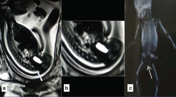
A 23-year-old pregnant lady presented for foetal MRI with antenatal foetal ultrasound scan showing absence of sacral vertebrae. Foetal MRI revealed abnormal morphology of the spine with loss of normal tapering of terminal vertebrae, missing sacral vertebrae and shortened spine [Table/Fig-1]. The conus was centrally positioned in spinal canal and seem to be terminated at the level of L1 vertebra, morphology of termination was wedge-shaped with abrupt termination. There was shortening of dural sac with smooth tapering. Rest of the spinal survey did not reveal significant point of note relating to open spinal defects.
a) Sagittal HASTE sequence and; b) Magnified view show absence of sacral vertebrae with loss of normal tapering of sacrum; c) Postnatal radiograph confirmed the absence of sacrum.
Correlative Ultrasonography (USG) revealed absence of sacrum, the shield like appearance of the fused iliac wings and the decreased interspace between the femoral heads. Lower limb screening was unremarkable for club foot. Urinary bladder was visualized; mild pelviectasis was noted in left kidney. Right kidney showed normal size of pelvis.
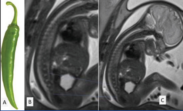
Ultrasound and foetal MRI findings were consistent with sacral agenesis, which was confirmed on post-natal radiograph [Table/Fig-1]. The normal tapering of the sacral vertebrae seen on foetal MRI resembles tapering of a chilli. This normal tapering is consistently visualised in all foetal MRIs and helps to ascertain the presence of sacrum in foetus (chilli sign) [Table/Fig-2]. Absence of this normal tapering seen on foetal MRI should lead to diagnosis of sacral agenesis.
Sagittal HASTE sequence shows normal tapering of sacrum resembling that of Chilli (a, b and c). This normal tapering of sacrum (chilli sign) is consistently visualised in all normal fetal MRI, and helps to rule out sacral agenesis.
Sacral agenesis is a rare developmental abnormality, observed more frequently in infants of diabetic mothers [1]. Sacral agenesis is classified into four types depending upon partial/complete agenesis, unilateral/bilateral absence of bony elements of sacral vertebrae and fusion of the iliac bones with lumbar vertebrae or each other (Renshaw classification) [2]. Classification of caudal regression syndrome is based on MRI and takes into account of embryological basis of development of spinal cord. Two types are classified based upon location and shape of conus medullaris [3]. Type 1 is characterised by abrupt spinal cord termination above L1 level. It is associated with sacral agenesis, bowel and bladder dysfunction. In type 2, there is low lying conus associated with tethering of filum terminale [4].
Conclusion
The normal tapering of sacrum is present in all foetal MRI, which mimics the shape of a chilli and helps in ruling out sacral agenesis.
[1]. Kumar Y, Gupta N, Hooda K, Sharma P, Sharma S, Kochar P, Caudal regression syndrome:a case series of a rare congenital anomalyPol J Radiol 2017 82:188-92. [Google Scholar]
[2]. Sharma S, Sharma V, Awasthi B, Sehgal M, Singla DA, Sacral agenesis with neurogenic bladder dysfunction—a case report and review of the literatureJ Clin Diagn Res 2015 9(6):RD08-RD09. [Google Scholar]
[3]. Pang D, Sacral agenesis and caudal spinal cord malformationsNeurosurgery 1993 32(5):755-79. [Google Scholar]
[4]. Negrete LM, Chung M, Carr SR, Tung GA, In utero diagnosis of caudal regression syndromeRadiol Case Rep 2015 10(1):1049 [Google Scholar]