Tiny Creature in Oral Cavity: A Case Report
Cheena Singh1, M Parvathi Devi2, Kamal Sagar3
1 Assistant Professor, Department of Oral Medicine and Radiology, Post Graduate Institute of Dental Sciences, Rohtak, Haryana, India.
2 Head, Department of Oral Medicine and Radiology, Teertnaker Mahaveer Dental College and Research Institute, Moradabad, Uttar Pradesh, India.
3 Demonstrator, Department of Periodontology, Post Graduate Institute of Dental Sciences, Rohtak, Haryana, India.
NAME, ADDRESS, E-MAIL ID OF THE CORRESPONDING AUTHOR: Dr. Cheena Singh, Assistant Professor, Department of Oral Medicine and Radiology, Post Graduate Institute of Dental Sciences, Rohtak-124001, Haryana, India.
E-mail: cheenasngh3@gmail.com
Oral cavity is nidus of much pathology. Some may arise due to altered eating habits such as meat, uncooked food etc. The parasitic infections arising from ingesting uncooked pork (larva of pork tapeworm) called as cysticercosis and being undiagnosed lead to “diagnostic dilemma”. Herein we report a case of cysticercosis involving the right dorsum of tongue along with review of literature.
Cysticercosis, Orofacial region, Tapeworm larva, Tongue
CASE REPORT
A 45-year-old male patient reported in Department of Oral Medicine and Radiology with chief complaint of swelling present on right upper surface of tongue since last 8-9 months. History of present illness revealed that initially the swelling was small now it has reached to the present size. Medical and family history were non contributory and there was no history of previous such swelling present to any site of face. Personal history revealed that patient was vegetarian and used to take 1-2 pegs of whisky every evening from past two years. On general physical examination, all the vital signs were within normal range.
Clinical examination of lesion revealed a well defined circular swelling present on right dorsum of tongue of approximately 1 cm x 1.5 cm, overlying mucosa appears to be normal. On palpation, swelling was soft to firm in consistency which was non tender and non fluctuant [Table/Fig-1].
Patient profile picture and swelling (white arrow).

Based on the history and clinical examination, cystic lesion involving right dorsum of tongue was given as provisional diagnosis with a differential diagnosis of soft tissue abscess.
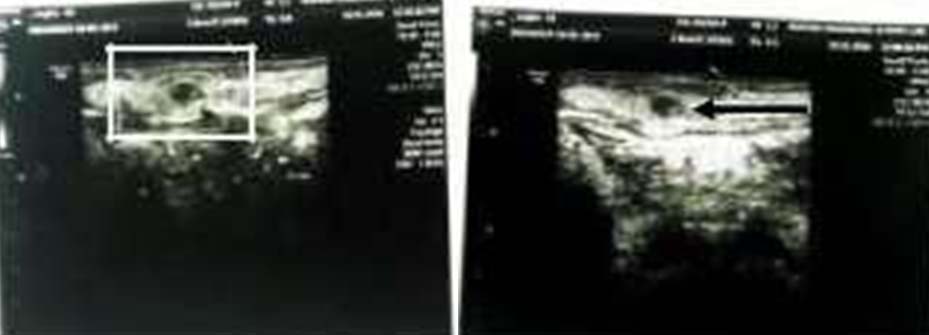
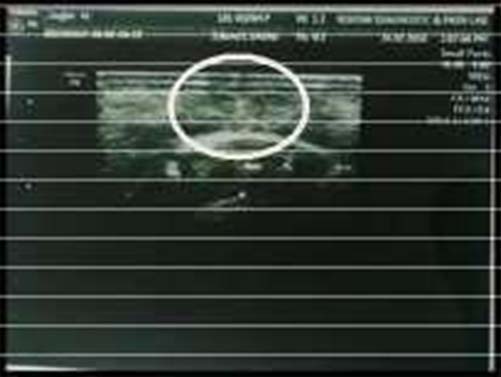
Aspiration (fine needle aspiration) of swelling was done yielding 0.2-0.5 ml of scanty material which showed deep blue nuclei in the background of fibrilar material. Smear also showed mixed inflammatory infiltrate showing possibility of cysticercosis. Patient was advised with various investigations, and on haematological investigations all the values were within normal limits except for the elevated ESR suggestive of parasitic infection. As the lesion was soft in consistency, so ultrasound of swelling using most latest version P8 4D was done which showed cyst of approximately 4 mm x 3 mm with a scolex (Arrow) at the point of complaint suggestive of cysticercosis [Table/Fig-2]. To rule out systemic involvement, ultrasound of abdomen was performed by PGIMS, Rohtak, Haryana, India (medical wing), but no systemic findings were found. No systemic and neural manifestations were reported in this patient thus CT and MRI were not performed.
Ultrasound (Preoperative) of swelling showing cyst with a scolex (Box and Arrow).
Based on the history, clinical findings and investigations, a final diagnosis of cysticercosis involving right dorsum of tongue was given.
Patient was prescribed Cap Albendazole 200 mg bid for 21 days. After five days he was reported with increase in pain at area of lesion. Then he was prescribed Tab Wysolone (prednisone) 20 mg for one week and recalled after one week for tapering of dose of tablet Wysolone to 10 mg. After 21 days of recalled, a remission of 60%-70% was noticed in lesion. Patient recalled after seven days and advised a postoperative ultrasonograph to assess the effectiveness of medications [Table/Fig-3].
Ultrasound (Postoperative) of swelling showing no hypodense areas with no scolex.
As the patient in the present case did not report with occurrence of cysticercosis at any other site or involvement of neurological symptoms, thus no additional treatment was prescribed except the periodic follow ups of every one month.
Discussion
Cysticercus is derived from Greek words Kystis = cyst and Kertos = tail because of their appearance [1]. It appears as soft, cystic to firm submucosal nodule which can be tender (symptomatic) and non tender (non-symptomatic) depending upon the inflammation initiated by cysticercus. The most common anatomical extraoral sites of cysticercosis occurrence are subcutaneous layers, brain, muscles, heart, liver, lungs, and peritoneum. The most commonly involved intraoral sites are buccal mucosa, lips and sometimes in tongue musculature [2].
Cysticercosis is an infection manifested by larval (cysticercus) stage of Taenia solium in the tissues of human body [3]. The definitive host is man and intermediate host is pig which harbours larval stage [4]. The adult worm lives in the small intestine of man. Usually one adult worm is present which lives for years having a length of about three meters with 1000 proglotids. The gravid segments have about 50,000 eggs in each segment The eggs hatch in small intestine, releasing oncospheres that penetrate the bowel mucosa and enter the bloodstream to reach various tissues where they develop to form a cysticercus cellulosae, which is the encysted larval form of T.Solium [5].
Although it can be diagnosed by first known conventional diagnostic modality which includes ultrasound where it appears round cyst with an eccentric ehcogenic protrusion from wall called as scolex as compared to MRI which is a costly advanced diagnostic modality. When multiple cyst calcifies it give a “starry night” appearance on computed tomography and multiple millet seed – shaped elliptical calcification in soft tissue on plain radiography [6].
Differential diagnosis of oral lesion depends on site involved. In the present case the differential diagnosis to be considered was mucocele, neurofibroma, vascular malformation, fibroma. Histopathological diagnosis is based on the recognition of cysticercus in the excised lesion. Viable cysticercus is usually present in excised tissue from the oral mucosa, breast, conjunctiva, subcutaneous tissues and other sites where presence of cysticercus arouses some concern and is readily accessible [1]. Thus, to prevent misdiagnosis proper clinical examination and relevant investigations should be done.
Patel K et al., stated that immunodetection can be performed by using Enzyme linked Immunosorbent Assay (ELISA) or Enzyme linked Immune Electrotransfer Blot (EITB) [5]. Although various treatment modality such as antihelminthics (including praziquantel) and surgical treatment are present for treating cysticercosis.
But in present study, Cap Albendazole was given along with Tab Wysolone as he complained of pain also in same lesion.
Pinswadi P and Charoensiri DJ [7] stated that cystecercosis should be treated with antihelminthic medication such as albendazole or prziquantel. In some cases, predenisolone should be given to prevent the inflammation due to cystecerci.
In present case, cysticercosis was reported in tongue musculature. Very few cases on tongue have been reported in the literature. Till date, only 35 cases have been reported in literature [1] and this one add to the present list.
Conclusion
Cysticercosis can be present extraorally and intraorally in human body. But in present article, a rare case of lingual cystecercosis have been diagnosed radiologically based on the sonographic appearance showing scolex which is pathogonomonic of cysticercosis. Proper clinical and radiological knowledge of this lesion is mandatory, so that a definitive diagnosis can be made with more confidence and thus the treatment should be started without any delay.
[1]. Koteeswaran G, Mangala G, Kotasthane DS, Tirou AT, Cysticercosis of tongue: Cytohistologic approach to diagnosisJournal of Oral and Maxillofacial Pathology: JOMFP 2013 17(3):480 [Google Scholar]
[2]. Elias FM, Martins MT, Foronda R, Jorge WA, de Araújo NS, Oral cystrcoicesis: case report and review of the literatureRev Inst Med trop S Paulo 2005 47(2):95-98. [Google Scholar]
[3]. Sharma P, Neupane S, Shrestha M, Dwivedi R, Paudel K, An ultrasonographic evaluation of solitary muscular and soft tissue cysticercosisKathmandu University Medical Journal 2010 8(2):257-60. [Google Scholar]
[4]. Dysanoor S, Pol J, A solitary facial nodular swelling - A case report of intramuscular cysticercosis in buccinator muscleAsian Pac J Trop Dis 2013 3(3):235-39. [Google Scholar]
[5]. Patel K, Shah M, Patel B, Doshi N, Subcutaneous oral cysticercosisNational Journal of Community Medicine 2011 2(2):311-13. [Google Scholar]
[6]. Boopathy Vijayaraghavan S, Sonographic appearances in cysticercosisJ Ultrasound Med 2004 23:423-27. [Google Scholar]
[7]. Pinswadi P, Charoensiri DJ, Cysticercosis in labial tissue. Case reportAust Dent J 1997 42(5):319-21. [Google Scholar]