Development of every individual is influenced by various factors like genetic, racial, nutritional, hormonal, environmental and climatic conditions [1,2]. According to Stewart RE and Barber TK the age of an individual can be determined by various methods like chronological age, biological age, morphologic age, skeletal age, dental age, circumpubertal age, behavioural age, mental age and self–concept age [2]. Chronological age is the most apparent and easily determined developmental age which is simply figured from child’s date of birth, but is not an accurate indicator of stage of development as it varies from individual to individual [2,3]. Hence, efforts were made to determine a child’s developmental age by using dental age (calcification of teeth) and skeletal age (skeletal maturation). Dental age can be estimated by using two principles like tooth emergence and tooth calcification of which the tooth calcification stages are considered more reliable indicator than tooth emergence, since it is less influenced by local and systemic factors [3-7]. Estimation of dental age was done based on calcification stages using Demirjian’s method. This method is most widely accepted because of its radiographic and schematic illustrations of tooth development and simplicity [4,8].
On the other hand, skeletal age is determined by recognizable stages of ossification and a characteristic pattern of progression of ossification of epiphyseal centres [9]. Fishman LS proposed the use of ossification centres seen in hand wrist radiographs and cervical vertebrae in lateral cephalogram [10]. However, its usage in pediatric patients is minimal due to various reasons like radiation exposure, high cost, huge equipment and the visibility of vertebra [10,11]. Hagg U and Taranger J proposed a method in which ossification of middle phalanx in the MP3 region was described as five stages of development namely MP3F, MP3FG, MP3G, MP3H, MP3I [11]. This method has many advantages over others such as low radiation exposure, high correlation with the six maturational stages of cervical vertebrae, no superimposition of bones or variations in posture and no need of special X-ray equipment. Abdel Khader HM first used intraoral periapical radiograph for assessing the reliability of the five stages of MP3 [12]. Later Rajagopal R and Kansal S modified MP3 method by adding MP3HI stage [13]. It has been well documented that the chronological age does not necessarily correlate with the maturational status of a child. However, high correlations exist between dental and skeletal age and these are more reliable and precise than chronological age in assessing the development of an individual [14]. Thus, the present study was aimed to correlate the chronological age, dental age and skeletal age in children between 6-14 years from Southeastern region of Andhra Pradesh, India.
Materials and Methods
In this cross-sectional study, a total of 900 patients in the age ranging from 6 to 14 years without sex predilection and who visited as outpatients to the Department of Paedodontics and Preventive Dentistry, Sibar Institute of Dental Sciences, Guntur, Andhra Pradesh, India, during December 2010 to June 2012 were screened. Well nourished children of Indian origin with pure ancestral background belonging to India and who require skeletal maturity assessment for orthodontic evaluation were selected for the study. The selected children presenting with all the seven permanent teeth on the left side of mandible which were evident radiographically, with no previous history of trauma and serious illness. Children with previous history of trauma to hand and wrist region, congenitally missing or extracted permanent teeth and aplasia of atleast two corresponding teeth bilaterally in mandible were excluded from the study. Premature, malnourished and syndromic children were also excluded from the study.
After attaining the Institutional Ethical Clearance, the parents/guardians of the screened patients were thoroughly explained regarding the study purpose and procedure. Out of the total 900 screened patients, only 100 children (48 males and 52 females) gave their consent to participate in the study. Standard radiation safety protocol was followed prior to the radiographic exposure. Two radiographs were made in each subject, a digital panoramic radiograph for determining the dental age by Demirjian method [4] and a digital radiograph for recording MP3 [Table/Fig-1,2]. The radiographs were numbered accordingly for future identification. The chronological age for all the subjects was calculated by substracting the date of birth from the date of radiographs made [3]. Digital Panoramic radiographs were taken using extraoral radiograph machine (SINORA) that uses exposure parameters of 8 MA, 71 kvp and 14.1 seconds. The seven permanent teeth on the left side of mandible were rated on a scale from A to H [4]. According to the criteria given by Demirjian for each stage and comparing the tooth diagrams [Table/Fig-3] with radiographs, the ratings were made. These ratings were converted into a score by using standard tables given by Demirjian for girls and boys separately. The obtained scores of all seven teeth were added together, which gives the total maturity score. This maturity score is directly converted into dental age applying Demirjian (1973) standardized tables for girls and boys [4]. The modified MP3 method in the present study was determined by MP3 radiograph, using digital radiography (SUNI), which uses exposure parameters of 8 MA, 70 kvp and 0.32 seconds [11,13].
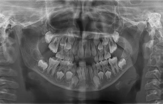
Digital Panoramic radiograph.
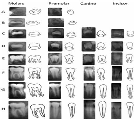
Demirjian tooth stages – comparison of radiographs with line drawing.
Courtesy: Liversidge HM [8]
Statistical Analysis
The values derived from the study were noted by a single examiner and statistically analyzed using SPSS version 17.0 (IBM, Chicago, IL, USA). Pearson’s and Spearman’s correlation tests were done to estimate the correlation between chronological, dental and skeletal ages among study population. To check reliability of the values obtained, 20 subjects were randomly selected and measured by a separate investigator who was unaware of the prior values. There was no significant variation in the values obtained by the separate investigator.
Results
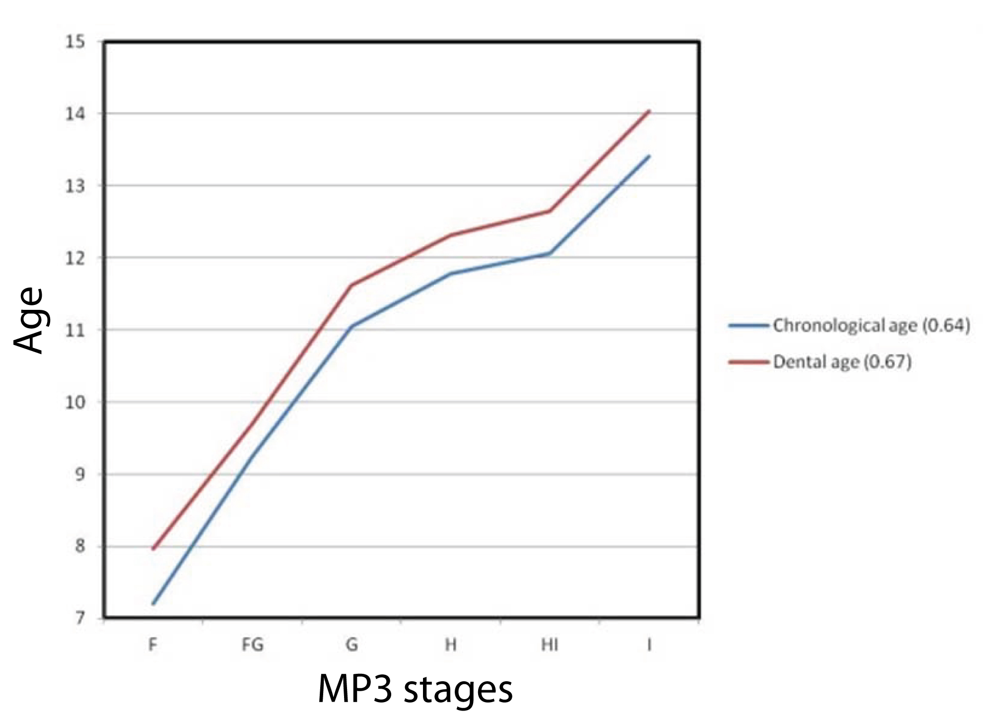
The results of the present study revealed statistical significance (p<0.05) between chronological age, dental age (Demirjian’s method) and skeletal stages of MP3 among all the subjects (48 males and 52 females). Males depicted significant positive correlation between chronological age, dental age and all stages of MP3 [Table/Fig-4]. The scenario was almost similar in females, except for a non-significant moderate correlation between chronological age and dental age in the H stage of ossification of middle phalanx in the MP3 region [Table/Fig-5]. Spearman’s correlation for the ossification stages of middle phalanx in the MP3 region among males with both the continuous variables of chronological age and dental age exhibited a strong positive correlation [Table/Fig-6] whereas the correlation found among females between these variables was not as high as that in males, but significant nevertheless [Table/Fig-7].
Correlation between chronological age and dental age among males at different stages of ossification of middle phalanx in the MP3 region.
| Stage | Mean chronologicalage (years) | Mean dentalage (years) | Correlationcoefficient (r-value) | p-value |
|---|
| F | 7.69±0.55 | 8.25±0.48 | 0.7 | 0.031 |
| FG | 10.01±1.51 | 10.62±1.49 | 0.63 | 0.039 |
| G | 11.37±0.38 | 11.75±0.21 | 0.68 | 0.05 |
| H | 12.30±0.43 | 12.99±0.63 | 0.712 | 0.028 |
| HI | 13.89±0.78 | 14.49±0.94 | 0.84 | 0.01 |
| I | 14.55±0.32 | 15.48±0.33 | 0.69 | 0.023 |
(Pearson’s correlation test; p ≤ 0.05 considered significant)
Correlation between chronological age and dental age among females at different stages of ossification of middle phalanx in the MP3 region.
| Stage | Mean chronologicalage (years) | Mean Dentalage (years) | Correlationcoefficient (r-value) | p-value |
|---|
| F | 7.21±0.53 | 7.96±0.24 | 0.69 | 0.05 |
| FG | 9.24±1.12 | 9.70±1.29 | 0.52 | 0.046 |
| G | 11.05±0.60 | 11.62±0.58 | 0.673 | 0.04 |
| H | 11.78±0.63 | 12.32±0.85 | 0.46 | 0.078 |
| HI | 12.06±1.03 | 12.65±1.07 | 0.681 | 0.03 |
| I | 13.41±0.83 | 14.04±0.88 | 0.54 | 0.027 |
(Pearson’s correlation test; p ≤ 0.05 considered significant)
Correlation of chronological age, dental age with skeletal age among males.
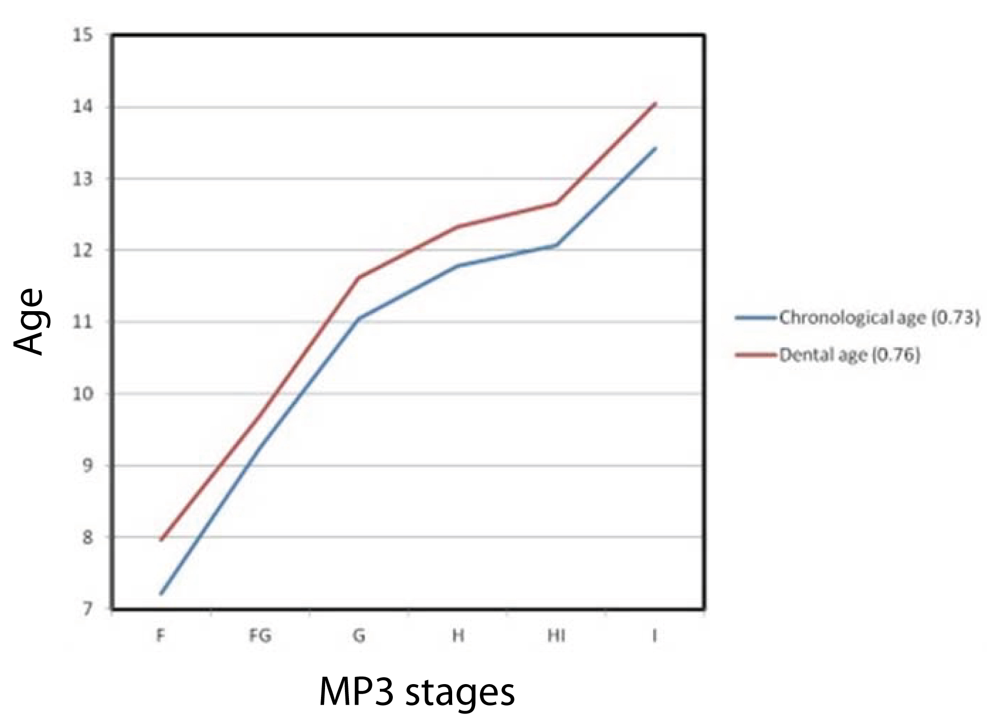
Correlation of chronological age, dental age with skeletal age among females.
Discussion
Determination of age can aid in the diagnosis of diseases that are age prevalent in the field of medicine and dentistry. Evaluation of age and maturational status of an individual will have a considerable influence on the diagnosis, management, execution and eventual outcome of the treatment which significantly contributes to the early correction of any skeletal and dental discrepancies in children.
In the present study, chronological age was assumed from the child’s date of birth which was obtained from the birth records. It was calculated by subtracting the birth date from the date on which radiographs were exposed. Ages were estimated on yearly basis and the decimal was taken to simplify statistical calculation [3]. Conversely, the dental age estimation was done using Demirjian method and skeletal age was calculated by modified MP3 method using the digital radiography [13].
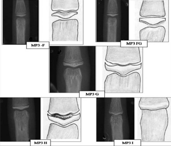
Skeletal age is considered more reliable and precise than chronologic age, as it provides visual inspection, appearance and ossification changes in shape and size of the bones. Fishman LS developed a system using four stages of bone maturation at six anatomic sites on the hand and wrist which are commonly used in clinics to assess the skeletal age [10], however there are concerns regarding the additional radiation exposure. So as to minimize the radiation exposure, the ossification of the distal phalanx of the first digit was used by Goto S et al., as an indicator to know the growth potential of that individual [15]. Abdel-Kader HM used these MP3 stages as seen on IOPA films [Table/Fig-8], for assessing skeletal maturity and later stated various advantages of digital radiography compared to that of conventional radiography like five times lower exposure time than conventional films and eliminating the darkroom procedures in addition to more clarity of the digital images [16]. This method also fulfils the principle of As Low As Reasonably Achievable (ALARA) [17].
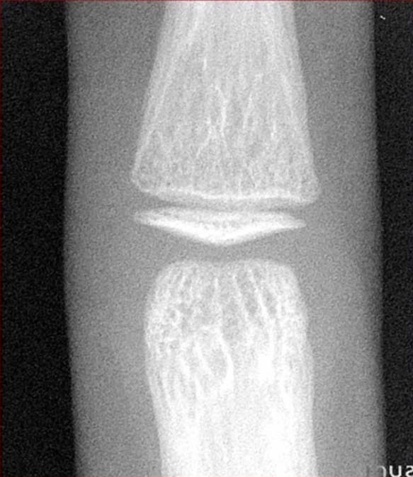
Radiographic interpretation of MP3 stages on IOPA.
Since there is literature debate regarding the relationship between skeletal and dental maturation, the current study is aimed at comparing the chronological, dental and skeletal age in children of 6-14 years. According to the study by Lewis AB and Gran SM [18] in Ohio children, there was no significant difference between skeletal and the dental maturation. On contrary, Sierra AM revealed high correlation (r=0.60) between dental and skeletal age in 153 orthodontically treated Caucasian children of age 8 to 12 years [19].
In the present study, the males exhibited high statistical significance (p<0.05) between chronological, dental and skeletal age. Similar results were reported by Hegde RJ and Sood PB where chronological age was compared with the dental age (Demirjian’s method) [20]. Uysal T et al., in a study noticed high correlation (p<0.01) between dental calcification stages and skeletal maturation stages [21], thus giving inference that the tooth calcification stages might be clinically useful as a maturity indicator for pubertal growth. In another study by Chaudary K et al., on a sample of 80 children aged between 8 to 14 years revealed a high correlation (p<0.01) between dental and skeletal age [22].
Correlation between chronological age, dental age and skeletal age in females was found to be statistically significant (p<0.05) except for the H stage of MP3 in the present study. The results of the present study revealed that females attained skeletal maturity earlier than chronological and dental age when compared to males. Studies conducted by Uysal T et al., on 500 Turkish children [21] and Soegiharto BM et al., on 2167 patients of Deuteromalay origin showed that on average girls mature faster than boys [23]. However, in studies by Chertkow S and Paul F on 140 children (93 girls and 47 boys) of Caucasoid origin [24] and Krailassiri S et al., on 261(39 male and 222 female) subjects of Thai origin found no statistical significant difference in both sexes [25]. The correlation between the dental and skeletal age in between males and females showed significant difference in HI and I stages and in remaining stages, there was no significance. In a study by Madhu S et al., the mean chronological ages observed in males and females of Mangalore population with respect to the stages of MP3 exhibited variance [26]. The reasons for the disparity could be the difference in the age group of the population, ethnic background, dietary pattern and the estimation method applied.
In the present study, dental age assessment done using Demirjian’s method showed high accuracy with an overestimation of 0.63 years in males and 0.59 years in females when applied to the population of age group 6-14 years age group. Similar results were obtained from the studies conducted by Hegde RJ and Sood PB, Koshy S and Tandon S, Prabhakar AR et al., who recorded an overestimation of 0.14 years, 3.04 years, 1.20 years for males and 0.04 years, 2.82 years, 0.90 years for females, respectively in different Indian populations [20,27,28]. This overestimation of dental age using Demirjian’s method may be due to the positive secular trend in growth and development, different nutritional and socioeconomic status, ethnic background and standard tables developed for French – Canadian population.
Even though various studies were carried out to correlate chronological, skeletal and dental ages, very few studies [3,6,9,25] were reported in the literature correlating the three parameters-chronological age, dental age using Demirjian method and modified MP3 method for skeletal age. Determination of chronological age is simple to calculate but not an accurate indicator of growth. Dental age assessment done by using Demirjian method has good reproducibility and reliability, but its applicability and universal acceptance still remains questionable in cases of anodontia, ethnic disparity and variations in the age estimation between geographical areas. The skeletal age assessment methods have a high accuracy in determining the growth status of an individual but the high radiation exposures is to be considered, therefore a simple digital radiograph of MP3 which satisfies all the criteria like low radiation exposure following ALARA principle, easier estimation, reliability, good reproducibility can be the choice.
Although strong correlation between skeletal and dental age is reported, yet it cannot be considered as the only reliable indicator in assessing the growth. Till date, no single accurate age assessment method had been proposed, however the dental calcification stages may be useful as an adjunct to skeletal maturity in assessing the growth of an individual.
Limitation
Limitations include the possible influence of external factors such as climate, environment, ethnic variation, socioeconomic status and internal factors like genetics, hormonal influences and nutritional deficiencies on growth of the child. The present study is limited to a certain group of population belonging to Southeastern region of Andhra Pradesh, India. Hence, further research is required on a larger sample with new population groups.
Conclusion
Chronologic age conveys only a rough approximation of the maturational status of a person, hence dental and skeletal ages had been explored as maturity indicators. The correlation between chronological, dental and skeletal age exhibited statistical significance in both males and females. Females attained maturity earlier than males in the present study population and the correlation between chronological and dental age in males and females showed statistical significance in HI and I stages of MP3. The proposed method of using digital radiography for assessing the skeletal age by MP3 method was simple with less radiation exposure and more reliable.
(Pearson’s correlation test; p ≤ 0.05 considered significant)
(Pearson’s correlation test; p ≤ 0.05 considered significant)