Introduction
The optimal way of restoring a non-vital tooth has long been a controversial matter. Decreased moisture content and loss of tissue either at radicular [1] or coronal level remains a major concern from biomechanical standpoint. One study [2] concluded no change in tissue moisture with loss of tooth vitality. Coronal and radicular destruction points out the highly conservative approach during endodontic and restorative procedures [3]. The significance of ferrule was established in the longevity of restoration of non vital tooth [4]. Thus, endodontically treated tooth that has been left with very less coronal tooth tissue requires dowel to retain the core. With the advancement of adhesive restorations, sometimes the dowel insertion procedure becomes unnecessary [5,6].
The intent of this article series was to assess the factors that may influence the successful restoration of root filled teeth with root canal dowels.
Review Method
The presented review type is literature review as characterized by methods used. This review includes comprehensive searching and typical narrative synthesis with thematic analysis. It lacks quality assessment of the literature search. Search strategy included a review of database using PubMed for the years 1990 to 2015. The search contained the following principal key terms: post/posts, dowel/dowels together with design, materials, retention, fracture resistance, biocompatibility, aesthetics, luting cements. Results were examined and repetitions were discarded. The search was kept limited to dental journals. Full papers were obtained wherever possible and where it is not possible to obtain the particular journal, abstracts were examined electronically. Inclusion criteria for the present review were, 1) any paper related to prefabricated/custom cast dowel/dowels; 2) only papers published in English language; 3) papers in peer reviewed journals. Additional references from papers were checked and included if met with inclusion criteria.
The original key terms resulted in 228 articles. Of these, which met with inclusion criteria for review was 51. The majority of studies were in vitro investigations. Few papers examined the physical properties of dowels.
Other published literature was case reports, RCT, FEA studies, SEM studies and systematic/non-systematic reviews.
When to Place Dowel?
Treatment decision with dowel depends on the amount of remaining tooth structure and functional demands that will be placed on the tooth [5]. Teeth with minimal remaining tooth structure provide decreased retention for the restoration and are at increased risk for fracture. As the remaining tooth structure decreases and functional forces increases, greater restorative control is needed.
The anterior teeth are not placed perpendicular to the occlusal plane and predominantly subjected to the labially inclined shear forces during function and therefore considered more prone for failure [7,8]. One study [9] has reported endodonticallv treated anterior teeth in which dowel was is not used for restoration, with greater fracture resistance, when compared with dowel-core crown restored maxillary anterior teeth. Dowel is not indicated, unless complete coverage restoration is required for aesthetic and functional reasons [4]. With maxillary central incisors and canines, amount of remaining coronal tooth structure, occlusion and function of the tooth are decisive.
For posterior tooth, indications are more precise. Posterior teeth are subjected to greater masticatory forces than anterior teeth. Endodontically treated posterior teeth should receive cuspal coverage as opposed to anterior teeth [10]. In most cases, they do not require a dowel. A dowel is indicated in posterior tooth only when there is extensive loss of coronal tooth structure or the tooth is to serve as an abutment for a removable or fixed partial denture. Premolars are mostly single rooted teeth with bulkier coronal structure. They are subjected to lateral forces more likely than molars during mastication. Hence, premolars require dowels more often than molars. Generally, dowel is to be avoided in buccal roots of maxillary molars and mesial roots of mandibular molars [11].
Remaining Tooth Anatomy after Endodontic Treatment
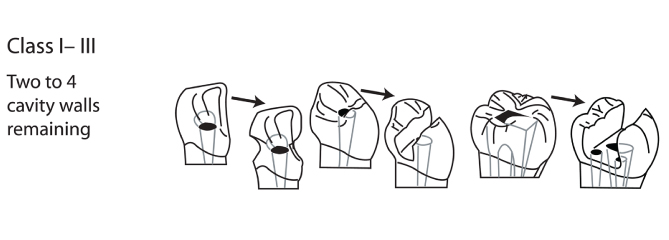
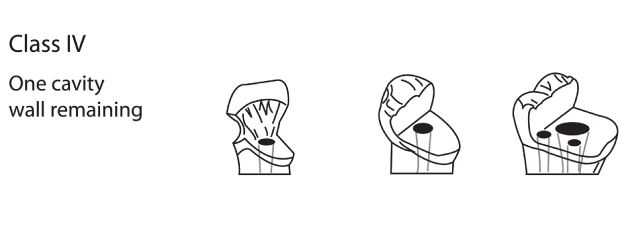
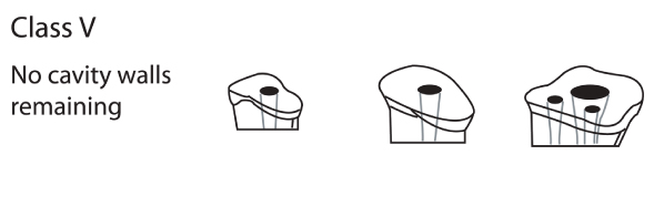
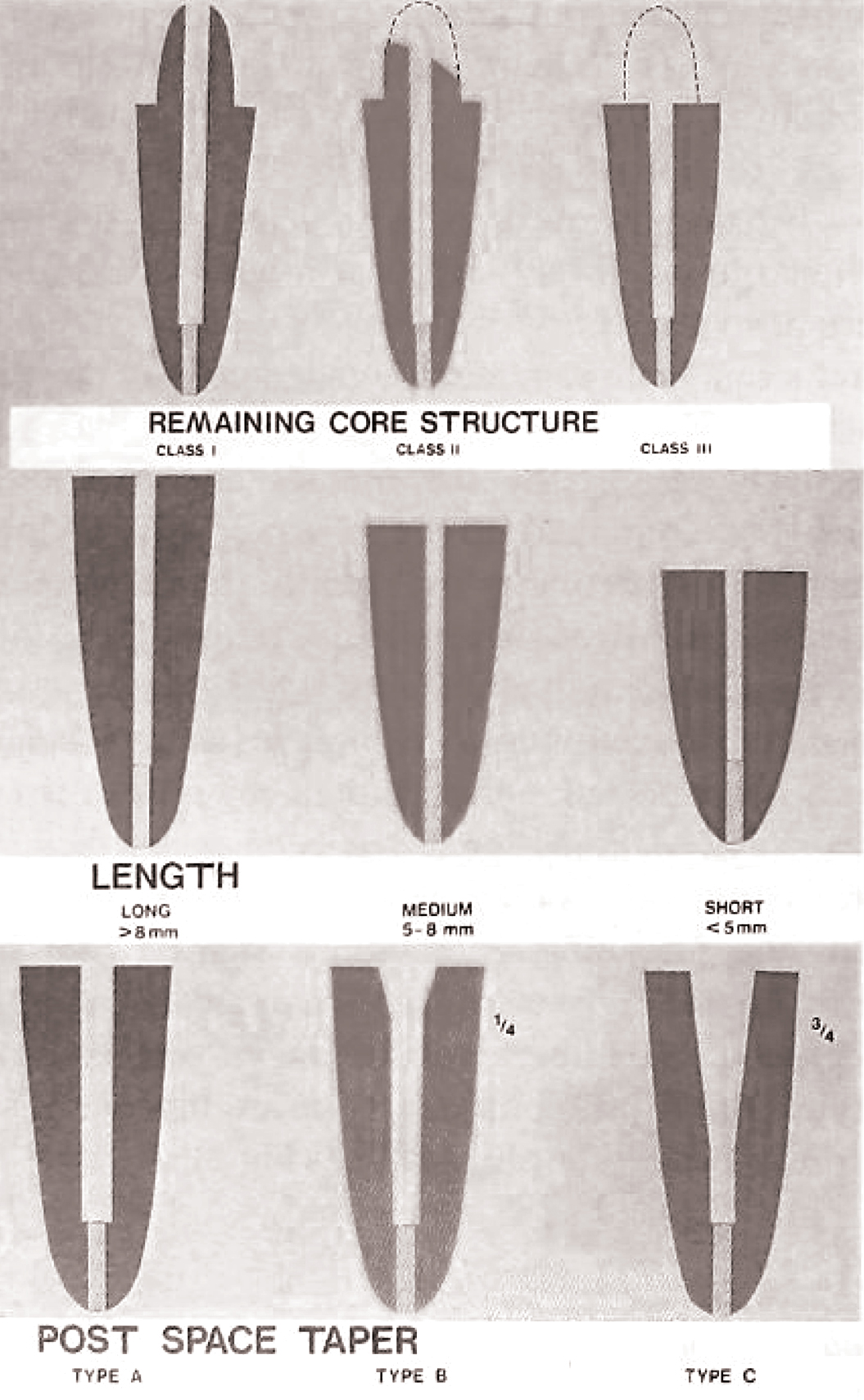
Root morphology and remaining coronal tooth structure affects the dowel selection. A consideration regarding the root size and length is important as improper dowel space preparation present the risk of apical or lateral root perforation. Radiographs serves to estimate root length, width and associated anatomic variations of the root canal [12]. Gutmann JL, [2] after reviewing the anatomic considerations found that roots of maxillary centrals, laterals and mandibular premolars have sufficient bulk to accommodate most dowel systems. Peroz I et al., [5] classified the tooth anatomy after endodontic treatment into five classes and assigned dowel indications to each class. Class I has four remaining cavity walls and it is not necessary to insert the dowel. Class II and III have two and three remaining cavity walls and do not require insertion of dowel [Table/Fig-1]. Class IV has one remaining cavity wall. Dowels are indicated in such cases of reduced remaining tooth structure [Table/Fig-2]. Class V has no remaining cavity wall and insertion of dowel is mandatory for core retention [Table/Fig-3]. Kurer H G [13] classified the single rooted pulpless teeth based on remaining coronal tooth structure, root length and taper of the dowel space [Table/Fig-4]. In Class I category, tooth has sufficient supragingival tooth structure for normal crown preparation. Class II has insufficient tooth structure for crown preparation but can be supplemented with suitable core without the need for dowel placement. Class III has no coronal tooth structure remaining. Length of the dowel has three variations long (8 mm), medium (5-8 mm), short (<5 mm). Taper was divided into three types. Type A has parallel dowel space, Type B has taper in coronal one third and Type C has taper in coronal three quarter. Length and taper are added as subclasses and described as ‘medium rooted Class III Type B’. Class IV is intraosseous fracture of root and in Class V periodontal disease is severe.
No dowel is needed in cases with at least two axial cavity walls remaining. A thickness of the cavity wall - 1 mm and a height of - 2 mm are preconditions. If these conditions cannot be fulfilled, the cavity wall must be considered as missing [5].
A dowel should be inserted if only one cavity wall is remaining. Fiber dowels are preferable in anterior teeth, but in posterior teeth, fiber or metal dowels can be used. The core can be made of composite or as a cast post and core. The definitive restorations should be crowns in anterior teeth and crowns, onlays or overlays in posterior teeth [5].
A post must be inserted if there is no cavity wall remaining [5].
Classification of pulpless teeth [13].
Dowel Space Preparation
Canal preparation for dowel placement is done by mechanical- rotary instruments, physical-heated instruments, chemical-solvents delivered with a hand file or combination of these methods. No method was found superior to other [14]. Heated endodontic plugger eliminates the possibility of inadvertent damage to the dentin but it is more time consuming. This method can be used to remove the guttapercha immediately after obturation without disturbing the apical seal. The procedure timing for preparing the dowel space is critical as it may displace residual filling material [14]. Epoxy resin based endodontic sealer requires an eight-hour setting time. A minimum 3-5 mm of gutta percha should remain at apical end to preserve the apical seal. Apical seal should be confirmed radiographically. At any location, dowel diameter should remain less than one third of the root diameter [11]. After removal of gutta percha, canal should be widened with appropriate endodontic reamers. Peeso-reamers and gates glidden drills are dowel preparation instruments. Gates glidden drill has football shape cutting head and often causes small concavities in root canal wall. Peeso-reamer is more cylindrical shaped. Accompanying twist drills can also be used to parallel the canal walls. The twist drills must not be forced into the canal or should not be used to remove the gutta percha from the canal [14]. Drills are end cutting instrument and can cause eccentricity by gauging the dentin or perforate the root. A recent study [15] assessed the adverse effect of endodontic sealers on retention of fibre posts. The study revealed traces of gutta perch and endodontic sealer on the walls of optimally prepared dowel spaces and concluded that over-preparation of dowel space helps to improve the retention.
Design Considerarations with Dowel
The endodontic dowels are broadly typed into custom cast dowel-core and prefabricated variety. The prefabricated dowels are classified as parallel, tapered or parallel- and- tapered combination based on their shapes [16]. They have also been classified as active or passive according to their surface characteristics. The active dowel has threads on the surface to be engaged into the root canal dentin whereas passive dowel is cemented with appropriate luting agent. Active dowels are more retentive and provides torsional resistance as they engage radicular tooth structure with threads but make root vulnerable to fracture. The indication for an active dowel is in a short canal in which there is an increased need for retention. Passive dowel largely depends on the close adaptation in the root canal for its retention.
Parallel sided dowel distributes functional load more passively along the dowel length except at the apex, where there is greater stress concentration as removal of tooth structure is more and definite apical seat of the dowel on dentin [17]. The tapered dowel preserves tooth structure at the apex of the dowel space but causes wedging effect and stress concentration at the coronal end [4]. Parallel- tapered dowel, a combination design of parallel sided dowel with tapered end, causes reduced stress concentration at the apex [16].
Threaded dowel exhibits unfavourable pattern of stress distribution during placement and during function. The concentration of stresses are seen at the dentinal thread interface. One half turn counter rotation reduces stress concentration at the interface but cannot reverse the damage that have already occurred while placement [16].
Torbjoner A et al., [18] compared failure rates and failure characteristics of tapered and parallel-sided dowels. They found the cumulative failure rate of tapered dowel was 15% higher than the failure rate for parallel-sided dowel (8%). Loss of retention was listed as the most frequent reason for failure for both types of dowels.
The serrated parallel dowel produces uniformly distributed stresses and protects the dentin. Tapered self-threaded dowel is not recommended as it causes stress and fracture very often [4].
Kayahan MB et al., [19] evaluated stress distribution in hollow and solid zirconia dowels. 3D FEA results revealed higher tensile stress in hollow design zirconia dowels during parallel and oblique load.
Dowel Material
Dowel materials can be divided into metallic and non metallic. Metallic dowels are either prefabricated or custom cast. Non metallic dowels include fiber dowels and ceramic/zirconia dowels. Fiber dowels are further classified into carbon, quartz or glass fiber reinforced epoxy resin dowels and polyethylene woven fiber embedded conventional composite resin dowels. Dowels are fabricated from a variety of materials and, therefore exhibit a variety of physical properties. To achieve optimum results, the material used for the dowel should possess modulus of elasticity similar to that of dentin and be biocompatible in the oral environment. Prefabricated metal dowels are very popular. Carbon fiber dowels [20,21], zirconia dowels [22] and composite material for dowels and cores [23] are also available. For largely intact tooth structure, all dowels that are in use today have sufficient strength and retention for clinical function. In various literature, dowel material has been evaluated in relation to retention and fracture resistance.
Among metal dowels, Stainless Steel (SS) is stiffer than titanium alloy, which is stiffer than pure titanium which is again stiffer than dentin. The use of metal dowel is justified by studies showing that the fracture resistance of teeth restored by metal dowel is superior to other systems [20]. However, in vitro studies [9,20,21,24] of uncrowned teeth have showed a greater risk of root fracture with metal dowels. Rough surface metal dowels were recommended to provide the best retention in the root canal [25-29]. The meta-analysis made by a systemic review of in vitro and in vivo studies [20] revealed no significant difference between cast and direct dowel and core systems that would justify recommending the use of one over the other.
In response to the need for a dowel that possesses good optical and biologic properties as well as compatible with an all ceramic crown, all ceramic dowel and core was developed in the late 1980s These dowels were made from fine grained tetragonal zirconium dioxide (Cerapost, Brasseler, Georgia; Cosmopost, Ivoclar-Vivadent, NY) [30-33] and was reported to possess high flexural strength [34] compared to the aluminium oxide ceramic material. In 1991, Kern M and Knode H [34] described slip casting technique for fabrication of aluminium oxide ceramic dowel. Copy milling is another technique for fabrication of glass-infiltered alumina ceramic dowel and core. Because of limited fracture toughness, glass-infiltered aluminium oxide ceramic (Inceram) dowels should be used in wide root canals without the crucial reduction of the circumferential dentin [30]. Aluminium oxide glass ceramic has improved esthetic properties due to increased translucency while zirconium dioxide ceramic has improved mechanical properties due to increase fracture strength. It is possible to combine both materials in single dowel and core unit by two piece technique or heat press technique [30]. It possesses high compressive strength, even though tensile strength is poor, hence when subjected to shear stresses, ceramic dowel itself fractures rather than root as in case of metal dowel [35]. Ziconia dowel is usually not preferred in bruxism patient due to high modulus of elasticity [36]. High elastic modulous of zirconia dowel causes stress transfer to less rigid dentin and predisposes to root fracture. The zirconia dowel is designed to be used with resin cement and composite core material. The IPS Empress core (Ivoclar) can be used to avoid less rigid, large composite resin core [22].
A carbon fiber dowel consists of bundles of stretched carbon fibers embedded in an epoxy matrix. The modulus of elasticity of carbon fiber dowel is similar to that of dentin [37]. The original version was inherently black, unesthetic and radiolucent. Esthetic version of this dowel has a quartz exterior that makes the dowel tooth colored. To overcome the disadvantages of carbon fiber dowel, glass fiber supported resin dowel systems were introduced in 1992. The dowel was composed of unidirectional glass fibers embedded in resin matrix that would improve the strength of the dowel without compromising the modulus of elasticity [38]. The retrospective in vivo study [39] concluded the superiority of fiber dowel to the conventional cast dowel and core systems after four years of clinical service. In cases of fractures, the fiber dowels produced more restorable fractures than other dowel materials [40-42]. Fiber dowel exhibits an additional advantage that it is readily retrievable after failure [40]. Soares LP et al., [43] concluded that FRC prostec (Ivoclar Vivadent Inc., NY) dowel would provide favourable response to masticatory forces as it had significantly higher flexure strength values than other types.
Fiber Reinforced Composite (FRC) resin dowel is made of woven polyethylene fiber ribbon that is coated with a dentin bonding agent and packed into the canal, where it is light polymerized [42] . Examples of fiber reinforced polymers are Ribbond (Ribbond Inc, Seattle, Washington), Fibrekore dowel System (Jeneric/Pentron, Connecticut). These all are categorized as non-metallic or esthetic dowels.
Types of Dowel
Endodontic dowel or dowels have been classified in various ways including preformed and custom cast, metallic and non metallic, esthetic and non esthetic. Dowel can also be classified as cemented/bonded dowel or threaded dowel.
Cemented dowels include custom cast dowel and a variety of prefabricated designs. There are over 100 different prefabricated dowel systems available. However, there are six basic commercial systems [14]. They are:
Tapered, smooth-sided dowels, such as Kerr Endopost dowels (Sybron-Kerr Inc., Romulus, Mich.).
Parallel, serrated and vented dowels, such as Whaledent Parapost dowels (Whaledent Int., New York, N.Y.).
Tapered, self-threaded dowels, such as Dentatus screws (Weissman Technology International, Inc., New York, N.Y.).
Parallel, threaded, split-shank dowels, such as FlexiPost dowels (Essential Dental Systems, S Hackensack, N.J.).
Parallel, threaded dowels; for example, Radix anchors (Maillefer/L. D. Caulk, Milford, Del.) or Kurer anchors (Teledyne Getz, Elk Grove, Ill.).
Carbon-fiber dowels, such as C-Post dowels (Bisco Dental Products, Itasco, Ill.) or Composipost dowels (RFF, Meylan, France).
Custom made dowels are indicated in moderate to severe loss of coronal tooth structure and in root canals with non-circular, elliptical cross section. Enlarging such canals for prefabricated dowel may lead to perforation in the apical area. In addition to that, cast dowel is recommended in following circumstances : 1. When core angle has to be changed in relation to the dowel; 2. when core retention is difficult on dowel head due to small size of the teeth such as mandibular incisor; 3. when multiple dowel core restorations are indicated in the same arch. The procedure can be completed in a more cost effective way by preparing multiple dowel spaces and making an impression. Prefabricated dowels are generally used in root canals with circular cross-section. Prefabricated tapered dowel is an ideal choice when there is an adequate canal length for axial retention. Prefabricated parallel dowel is recommended in need for increased retention and when preparation of parallel dowel space will not compromise the root integrity in apical third.
Dowel Diameter
Various investigators [44] have recommended different theories regarding the dowel diameter. Lloyd PM and Palik JF [45] have categorized these theories into conservationist, preservationist and proportionist. This proportionist approach was advocated to save sufficient tooth structure. Preservationist wherein the dowel should be surrounded by a minimum of 1 mm of sound dentin. Pilo R and Tamse A [44] advocated the conservationist approach wherein minimum canal preparation is done to conserve as much dentin as possible. Dowel diameter and the remaining dentin are identified as variables that influence the retention and fracture resistance of an endodontically treated tooth. Increasing the diameter of the dowel does not provide a significant increase in the retention of the dowel. Several studies observed that the greater the amount of remaining dentin, the better the fracture resistance. Tey KC and Lui JL [46] recommended the use of smaller FRC dowel rather than enlargement of dowel spaces to accurately fit larger FRC dowels. Dowel diameter must be controlled to preserve radicular dentin and to reduce the potential for perforations. The approach should be to thoroughly evaluate a current radiograph, limiting the width of the dowel to one third of the root diameter [11], limiting the dowel to a length of 7 mm apical to the canal orifice in maxillary and mandibular molars and placing the dowel in the palatal canal of the maxillary molar and in mandibular molars in distal canal.
Dowel Length
Dowel length has a significant effect on its retention. In most instances, retention increases as dowel length increases. The dowel length affects stress distribution and thereby fracture resistance in the canal. Several studies state that the dowel length should reach two-thirds of the entire root length [14]. Dowel placement beyond two-thirds of root depth found to increase stress in the apical region. Short dowels are potentially dangerous and evidenced with more incidence of root fracture [17]. Selection of dowel length also depends on the type of fixation. Dowel length is less important to fracture resistance when ferrule effect is present. Ferrule effect is usually provided by the crown bracing against the remaining supragingival tooth tissue. Apical root filling of four mm to six mm was found to be necessary for predictable apical seal [47,48]. Considering the need for both a sufficient ferrule effect and the remaining apical sealing, it is sometimes not possible to have the dowel length two-thirds of the root length. Nissan J et al., [49] showed that adhesive fixation can compensate for reduced retention of the shorter dowels. Several other guidelines for dowel length have been proposed but not widely followed. The dowel should equal the occluso-cervical dimension of the crown, should be longer than the crown, should end halfway between crest and apical end of the root should be as long as possible without disturbing the apical seal [14].
Dowel and Shape of the Root canal
Canal configuration aids in making a choice between a custom cast dowel and a prefabricated dowel [50]. Excessively flared root canals are managed most effectively with a custom dowel. Root canals with circular cross-section require prefabricated dowel. It is advantageous if selected dowel closely fits or conforms to the canal shape as less dentin removal is required, thereby enhancing fracture resistance of the tooth as well as retention of the dowel [4].
In a flared canal, large prefabricated parallel sided dowels by removing additional tooth structure in the apical area can also be used but not recommended as it may lead to root fracture in apical one third [12]. In addition, root reinforcement with composite is suggested for wide canals [12]. Composite resin is bonded to intracanal dentin. Intraradicular reinforcement increases the diameter of the root from external surface to the dowel interface. In addition to increasing the thickness of the root, it reduces the incidence of metal dowel show through the thin root canal dentin to darken the gingival tissue. Cast dowel and core restoration should be selected in a canal that requires extensive preparation due to the retention factor [51].
Discussion
Dowel and core provide predictable restorative options for endodontically treated teeth. This paper has summarized the available published literature on dowel systems with rigid inclusion criteria to limit the literature for review published in peer reviewed journals. Medline/PubMed, most commonly used database for searching the dental literature, was used for this paper also. The authors believe that this review provides a good discernment into the available published papers in this area [Table/Fig-5].
Characteristics of important studies.
| Study | Method | Outcomes |
|---|
| 1) Sorensen JA and Engelman MJ [4]. | Comparative analysis | Parallel sided dowel resulted in less fracture upon failure. Tapered dowel resulted in more apical and lingually directed fracture. |
| 2) Peroz I et al., [5]. | Review | Authors classified remaining tooth structure as to influence the indication for dowel core restoration. |
| 3) Mentink A et al., [7]. | Retrospective study | The study found 82% survival rate in anterior teeth restored with dowel and core after 10 yrs. |
| 4) Fernandes AS et al., [12]. | Review | The authors have discussed various factors influencing the selection of dowel-core assembly. |
| 5) Kurer HG [13]. | Review | The remaining coronal structure of single rooted pulpless tooth has been classified. |
| 6) Stockton LW [14]. | Review | Parallel sided passive dowel can be used successfully to restore most endodontically treated teeth. |
| 7) Othman HI et al., [15]. | Comparative analysis | Authors observed traces of gutta percha and resin sealer on the optimally prepared root canal walls. |
| 8) Ross RS et al., [16]. | Comparative study | Authors observed that when threaded dowel was allowed to contact at the end of the channel, higher strains resulted. |
| 9) Torbjorner A et al., [18]. | Retrospective study | Higher success rate for parallel sided serrated dowel compared to cast dowel. Most common reason for failure was loss of retention. |
| 10) Kayahan MB, et al., [19]. | FEA study | Finite element analysis of hollow and solid zirconia dowel revealed higher tensile stress values with hollow design during parallel and oblique loads. |
| 11) Dean JP et al., [24]. | Comparative in vitro study | Parallel and tapered dowel groups resulted in more incidence of root fractures compared to carbon fibre dowel group. |
| 12) Nergiz I et al., [25]. | Comparative in vitro study | Grit blasting the dowel and roughening the root canal wall resulted in increased resistance to dislodgement. |
| 13) Standlee JP and Caputo AA [28]. | Comparative study | Dowel with transverse serrated or crosshatched design were retained better in canal than threaded or longitudinal spiral design. |
| 14) Toksavul S [36]. | Case report | Bilateral maxillary lateral incisors were treated successfully with heat pressed ceramic crowns supported on zirconia dowel and core assembly. |
| 15) Usumez A et al., [38]. | Randomized controlled trial | Microleakage among 4 different dowel systems was evaluated : 1. Stainless steel (Parapost) 2. Glass fibre (Snowpost) 3. Polyethelyne composite (Ribbond) 4. Zirconia (Cosmopost). Snowpost and Ribbond showed the lowest coronal leakage. |
| 16) Ferrari M et al., [39]. | Retrospective clinical study | Authors concluded that composipost system was superior to the conventional systems at the end of 4 yrs of clinical service. |
| 17) Cormier CJ et al., [40] | An in vitro comparative study | Conventional dowel showed higher number of unrestorable root fracture and irretrievable dowel. The fiber dowel were readily retrievable after failure. |
| 18) Akkayan B and Gulmez T [41]. | An in vitro comparative study | Quartz fibre dowel exhibited higher resistance to fracture than titanium, glass and zirconium dowels. |
| 19) Soares LP et al., [43]. | An in vitro comparative study | FRC prostec fibre reinforced dowel demonstrated higher flexure strength compared to four other systems. |
| 20) Pilo R and Tamse A [44]. | Comparative study | Authors recommended minimal canal preparation to conserve as much dentin as possible. |
| 21) Lloyd PM and Palik JF [45]. | Review | Three distinct philosophies regarding dowel diameter has been identified from the literature : 1. Dowel with narrowest possible diameter 2. Dowel with apical diameter is 1/3 of the narrowest root diameter 3. Dowel with at least 1 mm of sound surrounding dentin. |
| 22) Tey KC and Lui JL [46]. | In vitro study | The study confirms that enlarging the dowel space may increase chances of unfavourable root fracture. |
| 23) Abramovitz L et al., [48]. | Comparative in vitro study | The remaining root canal fillings after dowel space preparation provides inferior seal compared to intact gutta percha filling. |
| 24) Nissan J et al., [49]. | Comparative in vitro study | Adhesive resin cement allowed the use of shorter dowel in the canal. |
The majority of literature is of in vitro investigations, evaluating the physical properties of the various dowel systems. The results produced by this in vitro investigations may not reliably guide the clinical practice. Fiber dowel system has been introduced with the claim by the manufacturer that the system has a Young’s modulus approximating that of natural tooth which should result in decreased stress concentration and allow homogeneous mechanical and chemical bonding to reinforce the tooth. Long term clinical data are not available to substantiate these claims. The review of the literature shows that there is a lack of in vitro and especially, clinical studies, correlating the amount of remaining tooth structure to the indication for dowels. The authors strongly feel the need of randomized control trials exploring the success of newer prefabricated dowel systems with varying degree of tooth loss and thereby do provide insight into the indications of dowel.
Conclusion
Dowels should be considered only when there is a need to retain the core, not with the intention of reinforcing an endodontically treated tooth. Dowel should be considered in cases of reduced remaining tooth structure such as with one cavity wall or without any remaining cavity wall. Dowel size should ensure 1 mm of surrounding dentin and 3 mm of apical root filling intact. Canal configuration and tooth location in the arch determine the selection between custom cast dowel and type of prefabricated dowel. The clinician should be knowledgeable in selecting the right type of dowel system to meet the mechanical and esthetic needs for each individual tooth.
[1]. Trope M, Ray HL Jr, Resistance to fracture of endodontically treated rootsOral Surg Oral Med Oral Pathol 1992 73:99-102. [Google Scholar]
[2]. Gutmann JL, The dentin-root complex: Anatomic and biologic considerations in restoring endodontically treated teethJ Prosthet Dent 1992 67:458-67. [Google Scholar]
[3]. Dietschi D, Duc O, Krejci I, Sadan A, Biomechanical considerations for the restoration of endodontically treated teeth: a systematic review of the literature, Part II (Evaluation of fatigue behavior, interfaces, and in vivo studies)Quintessence Int 2008 39:117-29. [Google Scholar]
[4]. Sorensen JA, Engelman MJ, Ferrule design and fracture resistance of endodontically treated teethJ Prosthet Dent 1990 63:529-36. [Google Scholar]
[5]. Peroz I, Blankenstein F, Lange KP, Naumann M, Restoring endodontically treated teeth with posts and cores-A reviewQuintessence Int 2005 36:737-46. [Google Scholar]
[6]. Christensen G, Posts: necessary or unnecessary?J Am Dent Assoc 1996 127:1522-24. [Google Scholar]
[7]. Mentink A, Meenwissen R, Kayser A, Mulder J, Survival rate and failure characteristics of the all metal post and core restorationJ Oral Rehabil 1993 20:455-61. [Google Scholar]
[8]. Milot P, Stein RS, Root fracture in endodontically treated teeth related to post selection and crown designJ Prosthet Dent 1992 68:428-35. [Google Scholar]
[9]. Sidoli NG, King PA, Setchel DJ, An in vitro evaluation of a carbon fiber based post and core systemJ Prosthet Dent 1997 78:05-09. [Google Scholar]
[10]. Costa LC, Pegoraro LF, Bonfante G, Influence of different metal restorations bonded with resin of fracture resistance of endodontically treated maxillary premolarsJ Prosthet Dent 1997 77:365-69. [Google Scholar]
[11]. Goodacre CJ, Spolnik KJ, The prosthodontic management of endodontically treated teeth: a literature review. Part III tooth preparation considerationsJ Prosthodont 1995 4:122-28. [Google Scholar]
[12]. Fernandes AS, Shetty S, Coutinho I, Factors determining post selection: A literature reviewJ Prosthet Dent 2003 90:556-62. [Google Scholar]
[13]. Kurer HG, The classification of single-rooted, pulpless teethQuintessence Int 1991 22:939-43. [Google Scholar]
[14]. Stockton LW, Factors affecting retention of post systems. A literature reviewJ Prosthet Dent 1999 81:380-85. [Google Scholar]
[15]. Othman HI, Elshinawy MI, Abdelaziz KM, Retention of fiber posts to the optimally and over-prepared dowel spacesJ Adv Prosthodont 2013 5:16-20. [Google Scholar]
[16]. Ross RS, Nicholls JI, Harrington GW, A comparison of stress generated during placement of five endodontic postsJ Endod 1991 17:450-56. [Google Scholar]
[17]. Isidor F, Brondum K, Ravnholt G, The influence of post length and crown ferrule length on the resistance to cyclic loading of bovine teeth with prefabricated titanium postsInt J Prosthodont 1999 12:78-82. [Google Scholar]
[18]. Torbjorner A, Karlsson S, Odman PA, Survival rate and failure characteristics for two post designsJ Prosthet Dent 1995 73:439-44. [Google Scholar]
[19]. Kayahan MB, Özkurt-Kayahan Z, Yay K, Kazazoğlu E, 3D FEA of hollow and solid design zirconia dental dowelsJ Prosthodont 2016 25(3):229-34. [Google Scholar]
[20]. Insua AM, Silva LD, Rilo B, Santhana U, Comparison of the fracture resistance of pulpless teeth restored with a cast post and core or carbon-fiber post with a composite coreJ Prosthet Dent 1998 80:527-32. [Google Scholar]
[21]. Isidor F, Odman P, Brondum K, Intermittent loading of teeth restored using prefabricated carbon fiber postsInt J Prosthodont 1996 9:131-36. [Google Scholar]
[22]. Sorensen JA, Mito WT, Rationale and clinical technique for esthetic restoration of endodontically treated teeth with the cosmopost and IPS Empress post systemQuintessence Dent Technol 1998 12:81-90. [Google Scholar]
[23]. Rudo DN, Karbhari BM, Physical behaviors of fiber reinforcement as applied to tooth stabilizationDent Clin North Am 1999 43:07-35. [Google Scholar]
[24]. Dean JP, Jeansonne BG, Sarkar N, An in vitro evaluation of a carbon fiber postJ Endod 1998 24:807-10. [Google Scholar]
[25]. Nergiz I, Schmage P, Platzer U, McMullan-Vogel CG, Effect of different surface textures on retentive strength of tapered postsJ Prosthet Dent 1997 78:451-57. [Google Scholar]
[26]. Cohen BI, Penugonda B, Pagnillo M, Schulman A, Hittelman E, Torsional resistance of crowns cemented to composite cores involving three stainless steel endodontic post designsJ Prosthet Dent 2000 84:38-42. [Google Scholar]
[27]. Rosin M, Fleissner P, Welk A, Steffen H, Heine B, The influence of surface configuration on the retention of posts designed for use with a cast-on techniqueQuintessence Int 2001 32:119-30. [Google Scholar]
[28]. Standlee JP, Caputo AA, Effect of surface design on retention of dowels cemented with a resinJ Prosthet Dent 1993 70:403-05. [Google Scholar]
[29]. Cohen BI, Pagnillo M, Newman I, Musikant BL, Deutsch AS, Retention of four endodontic posts cemented with composite resinGen Dent 2000 48:320-24. [Google Scholar]
[30]. Koutayas SO, Kern M, All-ceramic posts and cores: The state of the artQuintessence Int 1999 30:383-92. [Google Scholar]
[31]. Asmussen E, Peutzfeldt A, Heitmann T, Stiffness, elastic limit and strength of newer types of endodontic postsJ Dent 1999 27:275-78. [Google Scholar]
[32]. Kakehashi Y, Luthy H, Naef R, Wohlwend A, Scharer P, A new all ceramic post and core system: clinical, technical and in vitro resultsInt J Periodont Restor Dent 1998 18:586-91. [Google Scholar]
[33]. Rosentritt M, Fürer C, Behr M, Lang R, Handel G, Comparison of in vitro fracture strength of metallic and tooth coloured posts and coresJ Oral Rehabil 2000 27:595-98. [Google Scholar]
[34]. Kern M, Knode H, Posts and cores fabricated out of In-Ceram-direct and indirect methodsQuintessenz Zahntech 1991 17:917-25. [Google Scholar]
[35]. Shetty T, Bhat S, Shetty P, Aesthetic postmaterialsThe Journal of Indian Prosthodontic Society 2005 5:122-25. [Google Scholar]
[36]. Toksavul S, Esthetic enhancement of ceramic crowns with zirconia dowels and cores – A clinical reportJ Prosthet Dent 2004 92:116-19. [Google Scholar]
[37]. King PA, Setchell DJ, An in vitro evaluation of a prototype CFRC prefabricated post developed for the restoration of pulpless teethJ Oral Rehabil 1990 17:599-609. [Google Scholar]
[38]. Usumez A, Cobankara FK, Ozturk N, Eskitascioglu G, Belli S, Microleakage of endodontically treated teeth with different dowel systemsJ Prosthet Dent 2004 92:163-69. [Google Scholar]
[39]. Ferrari M, Vichi A, Garcia-Godoy F, Clinical evaluation of fiber-reinforced epoxy resin posts and cast post and coresAm J Dent 2000 13:15B-18B. [Google Scholar]
[40]. Cormier CJ, Burns DR, Moon P, In vitro comparison of the fracture resistance and failure mode of fiber, ceramic, and conventional post systems at various stages of restorationJ Prosthodont 2001 10:26-36. [Google Scholar]
[41]. Akkayan B, Gulmez T, Resistance to fracture of endodontically treated teeth restored with different post systemsJ Prosthet Dent 2002 87:431-37. [Google Scholar]
[42]. Sirimai S, Riis DN, Morgano SM, An in vitro study of the fracture resistance and the incidence of vertical root fracture of pulpless teeth restored with six post-and-core systemsJ Prosthet Dent 1999 81:262-69. [Google Scholar]
[43]. Soares LP, de Vasconcellos AB, da Silva AH, Sampaio EM, Vianna GA, The relationship between fibre post geometry and flexural properties: an assessment through a modified three-point bending testEur J Prosthodont Restor Dent 2010 18:158-62. [Google Scholar]
[44]. Pilo R, Tamse A, Residual dentin thickness in mandibular pre-molars prepared with gates glidden and ParaPost drillsJ Prosthet Dent 2000 83:617-23. [Google Scholar]
[45]. Lloyd PM, Palik JF, The philosophies of dowel diameter preparation: a literature reviewJ Prosthet Dent 1993 69:32-36. [Google Scholar]
[46]. Tey KC, Lui JL, The effect of glass fiber-reinforced epoxy resin dowel diameter on the fracture resistance of endodontically treated teethJ Prosthodont 2014 23:572-81. [Google Scholar]
[47]. Wu M-K, Pehlivan Y, Kontakiotis EG, Wesselink PR, Microleakage along apical root fillings and cemented postsJ Prosthet Dent 1998 79:264-69. [Google Scholar]
[48]. Abramovitz L, Lev R, Fuss Z, Metzger Z, The unpredictability of seal after post space preparation: A fluid transport studyJ Endod 2001 27:292-95. [Google Scholar]
[49]. Nissan J, Dimitry Y, Assif D, The use of reinforced composite resin cement as compensation for reduced post lengthJ Prosthet Dent 2001 86:304-08. [Google Scholar]
[50]. Smith CT, Schuman N, Restoration of endodontically treated teeth: a guide for the restorative dentistQuintessence Int 1997 28:457-62. [Google Scholar]
[51]. Cohen BI, Pagnillo MK, Condos S, Deutsch AS, Four different core materials measured for fracture strength in combination with five different designs of endodontic postsJ Prosthet Dent 1996 76:487-95. [Google Scholar]