In Vitro Evaluation of Enamel Microhardness after Application of Two Types of Fluoride Varnish
Fatemeh Molaasadolah1, Solmaz Eskandarion2, Atieh Ehsani3, Meysam Sanginan4
1 Assistant Professor, Department of Paediatric Dentistry, Shahid Beheshti University of Medical Science, Tehran, Iran.
2 Assistant Professor, Department of Dental Material, Shahid Beheshti University of Medical Science, Tehran, Iran.
3 Resident, Department of Paediatric Dentistry, Shahid Beheshti University of Medical Science, Tehran, Iran.
4 Dentist, Tehran, Iran.
NAME, ADDRESS, E-MAIL ID OF THE CORRESPONDING AUTHOR: Dr. Atieh Ehsani, Resident, Department of Paediatric Dentistry, Shahid Beheshti University of Medical Science, School of Dentistry, Daneshjoo Sqr., Blvd Evin, Shahid Chamran Highway, Postal code: 1983963113, Tehran, Iran.
E-mail: a.ehsani645@yahoo.com
Introduction
Use of fluoride compounds is one of the most effective ways of preventing decay and among these varnishes have high acceptance among different fluoride products.
Aim
Hence, the aim of this research was to evaluate the micro-hardness of tooth enamel after the usage of two different commercial products of fluoride varnish.
Materials and Methods
This in vitro experimental study was performed on 51 extracted premolar teeth. The teeth were divided randomly into three 17-membered groups. The first group received Duraflor varnish, the second group received Ariadent Iranian varnish while the third group received no treatment. Micro hardness of tooth enamel was measured utilizing Vickers method before and after the use of fluoride varnish. ANOVA, Tukey, and Wilcoxon statistical tests were utilized for statistical analysis of data.
Results
The comparison of mean change in micro hardness before and after the use of fluoride showed that increase in micro hardness in Duraflor varnish and Ariadent varnish group was significant when compared to control group (p<0.05) but no significant statistical difference was observed in terms of mean of micro hardness after intervention between two groups of Duraflor varnish and Ariadent varnish (p>0.05).
Conclusion
According to the findings, the use of fluoride significantly increased the enamel micro hardness which did not show a significant difference between two groups of Duraflor varnish and Ariadent varnish.
Dental enamel, Fluorides, Hardness test
Introduction
Enamel has a protective role for tooth structure against external factors but this structure suffers from changes or irreversible damages against some factors like acidic environment due to bacterial activity [1]. Dental caries occurs due to imbalance in the dynamic process of demineralization and remineralization of enamel. Adequate stable minerals in the saliva is one of the most important factor in enamel remineralization and enamel would be restored by minerals in the saliva or dental plaque in these conditions [2,3].
Despite the current progress, tooth decay continues to be a major problem in most communities [4]. As we know, prevention is the best and most effective method against tooth decay and cost-effective method when compared to the cost of treatment of caries [5]. Fluoride has been known as the most effective material in the prevention of tooth decay which is available in different forms [6]. Fluoride varnish (5%) is often preferred by most dentists because of the ease and speed of use, lower risk of being swallowed and patient preference [7].
The surface hardness of enamel refers to dental resistance against scratches, abrasion, and indentation as well as resistance against permanent curvature and deformation at the time of force exertion [8]. Microhardness test is widely utilized to evaluate tooth hardness [9]. The purpose of this in vitro study was to compare enamel Micro-hardness after using two types of fluoride varnishes.
Materials and Methods
This in vitro experimental study was carried on 51 premolar teeth which were extracted due to orthodontic purposes. The teeth that were included in the study did not have any caries, hypocalcification, erosion, or cracks. Evaluation was performed according to WHO criteria [10]. Teeth were washed in antiseptic solution of sodium hypochlorite after being extracted and then tooth samples were cleared using pumice paste without fluoride by Air motor (W and H Angel). Thereafter, they were examined by stereo microscope (Cartoon Optimal Industries Ltd, SCW-X model, made in Thailand) at 40X magnification. Square label with dimensions of 4×2 mm was placed on the buccal surfaces of teeth. All the remaining surfaces of the teeth were covered with transparent self cured acrylic (Acropars, made in Iran) to have same contact surface in all teeth regardless of shape, size or the groups. Thereafter, the primary microhardness of teeth was measured and recorded by applying 50 gm force [11] on each sample by Vickers Hardness Measuring device. In this step, the samples were divided randomly (using sequentially numbered containers) into three 17 membered groups. The first group was treated with 5% (Duraflor) varnish (G1) and samples of the second group were treated with 5% (Ariadent) varnish (PREVENTA) (G2). The third group was considered as the control group. Varnishes were applied after ensuring the dryness of enamel surface in two layers by special brushes. Samples were separately placed in distilled water in a glass container after drying of varnish and were placed for 24 hours in incubator (Pars Azma Company) at 37°C with the control group. The control group did not receive any fluoride treatment. Varnishes were slowly cleaned from teeth surface with periodontal curette after 24 hours to synchronize the loss [12]. The samples were entered into pH cycle so as to create laboratory conditions similar to the mouth. Each cycle was performed for one day (24 hours). Initially, samples were placed separately in a mineralization solution with 2.2 mM CaCl2, 2.2 mM NaH2PO4, 0.05 M acetic acid, adjusted with 1 ml KOH, 15 ml per tooth at pH = 4.5 for three hours and then immersed in distilled water for 30 minutes. Thereafter, samples were placed in a remineralization solution with 1.5 mM CaCl2, 0.9 mM NaHPO4, KCl 0.15 mM; 15 ml per tooth and pH = 7.0 for 20 hours and were again washed and immersed in distilled water for 30 minutes. This cycle was repeated for 10 times (10 days) [7,13]. After the end of the pH cycling microhardness of enamel was measured by an investigator who was not aware of the groups the specimens belonged to it.
Statistical Analysis
The results obtained were statistically analysed. Analysis of variance (ANOVA) was utilized for comparison of enamel Microhardness of three groups. Tukey test was utilized for pairwise comparison between groups and Wilcoxon Test utilized for comparison of enamel microhardness of each group before and after treatment. SPSS version 18.0 software was utilized for comparison of microhardness quantitative variable.
Results
The effects of Iranian and England fluoride varnish on enamel micro hardness of 51 premolar teeth were studied and compared in this research. Results obtained from the evaluation of enamel micro hardness before and after the application of fluoride in groups are fully explained in [Table/Fig-1].
The results of ANOVA between groups before and after intervention.
| Hardness | Groups | N | Mean | SD | DF(Between Groups) | F | p-value |
|---|
| Before intervention | Varnish (Duraflor) | 17 | 230.2 | 46.6 | 2 | 0.228 | 0.779(NS) |
| Iranian varnish | 17 | 230.8 | 34.5 |
| After intervention | Varnish (Duraflor) | 17 | 282.7 | 48.7 | 2 | 12.270 | 0.001(S) |
| Iranian varnish | 17 | 255.1 | 33.3 |
NS-Non Significant, S-Significant
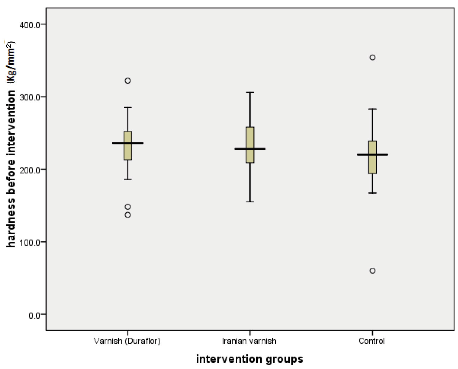
Results of ANOVA show that there was no significant difference in the average microhardness between groups before the intervention. In other words, variable values of microhardness among three groups before the intervention were homogeneous [Table/Fig-2].
Microhardness values of the three groups before the intervention.
Moreover, the results of enamel microhardness values after the application of fluoride in evaluated groups showed that there was a significant difference regarding the means of microhardness between some groups (p<0.05) [Table/Fig-3].
Microhardness values of the three groups after the intervention.
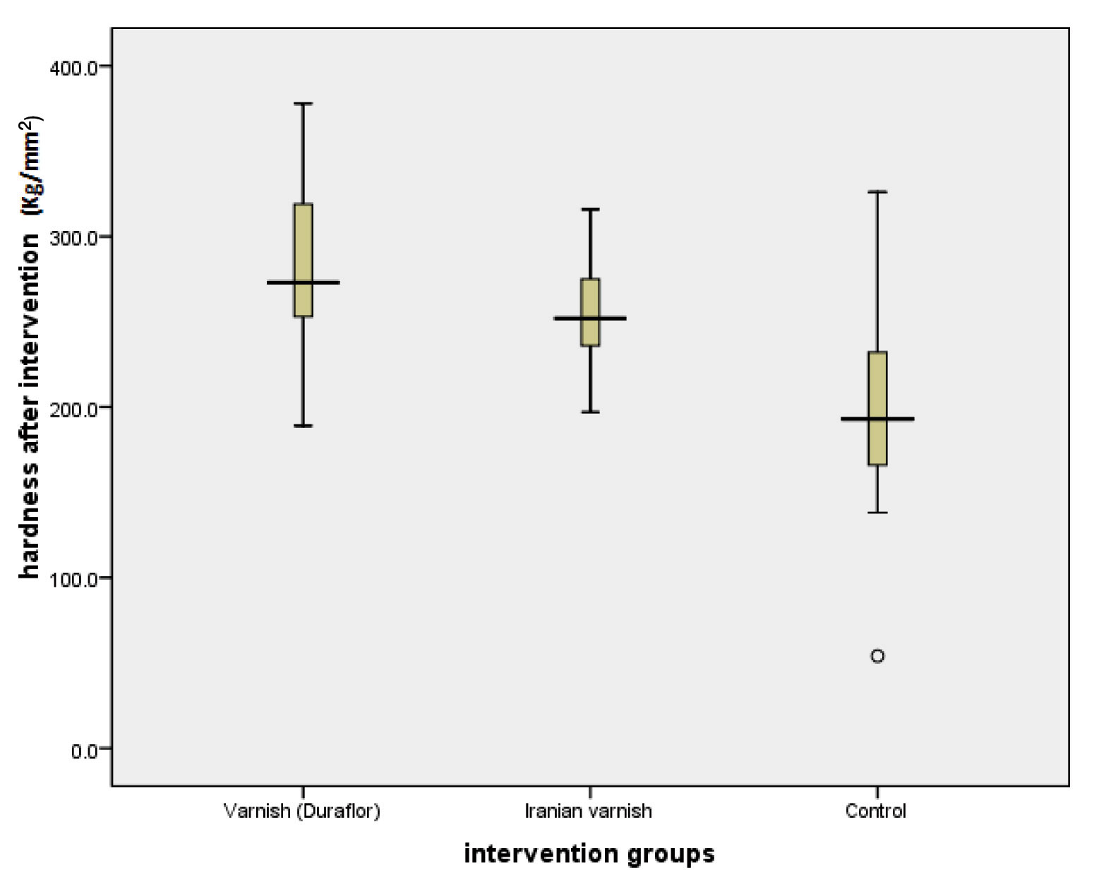
In other words, a significant difference was observed between the results of enamel micro hardness of two groups (G1 and G2) and results of control group. However, the value for Duraflor varnish was slightly higher than Ariadent varnish in evaluations carried out using Tukey test, but this difference was not significant in terms of average microhardness after intervention between Duraflor varnish and Ariadent varnish.
The evaluation was also performed using single sample Wilcoxon test which showed that the average microhardness was significantly increased after intervention in groups of Duraflor varnish as well as Ariadent varnish (273.7 vs. 236.3; p-value = 0.001, 252.1 vs. 228.3; p-value = 0.001 respectively) but average microhardness was significantly reduced in the control group (193.3 vs. 220.1; p-value = 0.001) [Table/Fig-4].
The results of Wilcoxon test in groups before and after intervention.
| Groups | Intervention | N | Mean | Median | Std. Deviation | Minimum | Maximum | p-value |
|---|
| Duraflor | hardness before intervention | 17 | 230.2 | 236.3 | 46.6 | 137.8 | 322.8 | 0.001 |
| hardness after intervention | 17 | 282.7 | 273.7 | 48.7 | 189.5 | 378.6 |
| Ariadent | hardness before intervention | 17 | 230.8 | 228.3 | 34.5 | 155.1 | 306.5 | 0.001 |
| hardness after intervention | 17 | 255.1 | 252.1 | 33.3 | 198.0 | 316.0 |
| Control | hardness before intervention | 17 | 220.6 | 220.1 | 62.3 | 60.3 | 354.4 | 0.001 |
| hardness after intervention | 17 | 200.2 | 193.3 | 61.9 | 55.0 | 326.8 |
Discussion
In this study, microhardness of tooth enamel after utilizing two types of varnish was evaluated and compared in vitro using Vickers Hardness Measuring method after laboratory simulation of oral environment. As mentioned earlier, pH cycling method by creating acidic challenges greatly helps in the simulation process of the mouth environment in the laboratory [14-16]. Nevertheless, 100% simulation cannot be expected because of the important and significant role of factors associated with remineralization process such as speed and flow rate of saliva and composition and buffering capacity of saliva [17,18].
Since the surface layer of enamel has a decisive role in the process of decay, evaluation of changes in this area has great importance. The calculation of surface’s micro hardness is a suitable method which was carried out using Vickers Hardness Measuring method. The advantage of this method is high accuracy and quantitative measurement capability [19]. In this method, there is the possibility of applying force with different sizes and the possibility of re-measurement of hardness of specimens within a specific time. However, the location of evaluation is not exactly the same as the previous point but in each group, hardness of each point can be considered as a symbol of the hardness of enamel surface because of the extreme proximity of evaluated points as well as the lack of significant differences between microhardness of initial points [20]. The values of surface micro hardness obtained in the present study were reported to be totally in the range of 227.2 kg/mm2 before the application of fluoride which is near to the range of reported microhardness for normal tissue of enamel [21]. The present study which was designed with the aim of evaluating effects of two types of fluoride varnish showed that there was a significant increase in enamel microhardness in both groups while this increase was not observed in the control group. Similar to the results obtained in this study, Nalbantgil D et al., concluded that microhardness after the application of fluoride varnish was more than the control group [22]. Sh P et al., compared the enamel surface microhardness after topical application of neutral sodium fluoride (NaF) and organic fluorides in the form of amine fluoride (AmF) solutions [23]. They concluded that since Fluoride augments the remineralization process by increasing the growth of demineralized enamel crystals, AmF compounds show a substantial increase in enamel micro hardness when compared to NaF. Dionysopoulos D et al., evaluated effect of fluoride treatments on bleached enamel Microhardness and surface morphology [24]. They concluded that the topical fluoride treatments (0.05% NaF daily, 0.2% NaF weekly and 5% NaF final topical fluoridation) after bleaching significantly enhanced the surface microhardness of the enamel. Gatti A et al., also concluded that toothpaste containing high concentrations of fluoride (1100 PPM) and fluoride varnish combined with toothpaste significantly reduced enamel demineralization compared to the negative control group [13]. The difference between microhardness was also observed in enamel before and after the application of fluoride varnish in the study of Navabi B et al., and Maia LC et al., stated that frequent utilization of fluoride toothpaste has a greater effect on Microhardness of enamel surface fluoride intake compared to when combining it with fluoride varnish [25,26]. In a study carried out by Tavassoli-Hojjati S et al., Iranian APF gel (Kimia) was able to significantly increase microhardness of tooth enamel compared to control group which is similar to the present study [27], but in a study of Biria MJ et al., unlike in the present study, fluoride gel (Sina) was not significantly different from the control group in terms of fluoride intake [28]. Although, Duraflor varnish increased the enamel microhardness slightly higher than Ariadent varnish in this study but there was no significant difference between the average microhardness after the intervention between two types of varnish (p<0.05). The study of Tavassoli-Hojjati S et al., also stated the same conclusion in relation to Iranian and foreign APF gel [27].
Lee YE et al., examined the effects of three types of topical fluoride treatment (solutions of NaF 2%, gel APF, cavity shield varnish) on remineralization of enamel caries lesions and concluded that fluoride treatment regimens do not have significant differences in the uptake of fluoride and reducing fluorescence lesion area [29].
These results indicate that two the types of varnish utilized in this study acted similarly in terms of increasing the enamel microhardness and it is suitable to use methods in evaluation of other aspects of the application of these varnishes.
As we know, remineralisation seen in vitro circumstances may be reasonable variable when compared to changes occurring under the clinical conditions in the oral cavity. Therefore, direct extrapolations to clinical situations must be executed discreetly.
Limitation
Since this study was accomplished in laboratory environment, it is not possible to reconstruct the real mouth characteristics. It is better to accomplish same studies in the mouth environment.
Conclusion
Within the limitation of the study, we conclude that there was a significant increase in tooth enamel micro hardness after the application of fluoride varnish. However, there was no significant difference between Ariadent varnish and Duraflor fluoride varnish.
NS-Non Significant, S-Significant
[1]. Kunin AA, Evdokimova AY, Moiseeva NS, Age-related differences of tooth enamel morphochemistry in health and dental cariesEPMA J 2015 6(1):3 [Google Scholar]
[2]. Mirkarimi M, Eskandarion S, Bargrizan M, Delazar A, Kharazifard MJ, Remineralization of artificial caries in primary teeth by grape seed extract: An in vitro studyJ Dent Res Dent Clin Dent Prospects 2013 7(4):206-10. [Google Scholar]
[3]. Peters MC, Strategies for noninvasive demineralized tissue repairDent Clin North Am 2010 54(3):507-25. [Google Scholar]
[4]. Reynolds EC, Calcium phosphate-based remineralization systems: Scientific evidence?Aust Dent J 2008 53(3):268-73. [Google Scholar]
[5]. Ramos-Gomez FJ, Crystal YO, Domejean S, Featherstone JD, Minimal intervention dentistry: Part 3. Paediatric dental care—prevention and management protocols using caries risk assessment for infants and young childrenBr Dent J 2012 213(10):501-08. [Google Scholar]
[6]. Vongsavan K, Surarit R, Rirattanapong P, The combined effect of xylitol and fluoride in varnish on bovine teeth surface microhardnessSoutheast Asian J Trop Med Public Health 2014 45(2):505-10. [Google Scholar]
[7]. AlAmoudi SA, Pani SC, AlOmari M, The effect of the addition of tricalcium phosphate to 5% sodium fluoride varnishes on the microhardness of enamel of primary teethInt J Dent 2013 2013:486358 [Google Scholar]
[8]. Westerman GH, Ellis RW, Latta MA, Powell GL, An in vitro study of enamel surface microhardness following argon laser irradiation and acidulated phosphatefluoride treatmentPediatr Dent 2003 25(5):497-500. [Google Scholar]
[9]. Chuenarrom C, Benjakul P, Daosodsai P, Effect of indentation load and time on knoop and vickers microhardness tests for enamel and dentinMaterials Research 2009 12(4):473-76. [Google Scholar]
[10]. Mendes FM, Braga MM, Oliveira LB, Antunes JL, Ardenghi TM, Bönecker M, Discriminant validity of the International Caries Detection and Assessment System (ICDAS) and comparability with World Health Organization criteria in a cross-sectional studyCommunity Dent Oral Epidemiol 2010 38(5):398-407. [Google Scholar]
[11]. Haghgou HR, Haghgoo R, Asdollah FM, Comparison of the microhardness of primary and permanent teeth after immersion in two types of carbonated beveragesJ Int Soc Prev Community Dent 2016 6(4):344-48. [Google Scholar]
[12]. Hong L, Watkins CA, Ettinger RL, Wefel JS, Effect of topical fluoride and fluoride varnish on in vitro root surface lesionsAm J Dent 2005 18(3):182-87. [Google Scholar]
[13]. Gatti A, Camargo LB, Imparato JCP, Mendes FM, Raggio DP, Combination effect of fluoride dentifrices and varnish on deciduous enamel demineralizationBraz Oral Res 2011 25(5):433-38. [Google Scholar]
[14]. Harris NO, Garcia-Godoy F, Nielsen Nathe C, Primary Preventive Dentistry 2014 8th edChap 1,5,14,15: 6,54,224-229,252-255 [Google Scholar]
[15]. Casals E, Boukpessi T, McQueen CM, Eversole SL, Faller RV, Anticaries potential of commercial dentifrices as determined by fluoridation and remineralization efficiencyJ Contemp Dent Pract 2007 8(7):1-10. [Google Scholar]
[16]. Ten Cate JM, Duijsters PP, Alternating demineralization and remineralization of artificial enamel lesionsCaries Res 1982 16(3):201-10. [Google Scholar]
[17]. Damato FA, Strang R, Stephen KW, Effect of fluoride concentration on remineralization of carious enamel: An in vitro pH-cycling studyCaries Res 1990 24(3):174-80. [Google Scholar]
[18]. Marsh PD, Microbiologic aspects of dental plaque and dental cariesDent Clin North Am 1999 43(4):599-614. [Google Scholar]
[19]. Rehder Neto FC, Maeda FA, Turssi CP, Serra MC, Potential agents to control enamel caries-like lesionsJ Dent 2009 37(10):786-90. [Google Scholar]
[20]. Ryge G, Foley DE, Fairhurst CW, Micro-indentation HardnessJ Dent Res 1961 40:1116-26. [Google Scholar]
[21]. Gutiérrez-Salaza MP, Reyes-Gasga J, Microhardness and chemical composition of human toothMat Res 2003 6(3):367-73. [Google Scholar]
[22]. Nalbantgil D, Oztoprak MO, Cakan DG, Bozkurt K, Arun T, Prevention of demineralization around orthodontic brackets using two different fluoride varnishesEur J Dent 2013 7(1):41-47. [Google Scholar]
[23]. Sh P, Raghu R, Shetty A, Gautham P, Reddy S, Srinivasan R, Effect of organic versus inorganic fluoride on enamel microhardness: An in vitro studyJ Conserv Dent 2013 16(3):203-07. [Google Scholar]
[24]. Dionysopoulos D, Koliniotou-Koumpia E, Tolidis K, Gerasimou P, Effect of fluoride treatments on bleached enamel microhardness and surface morphologyOral Health Prev Dent 2017 15(2):169-75. [Google Scholar]
[25]. Navabi B, Ansari G, Khan Z, Kheirieh P, Najafi B, Fluoride uptake level of the enamel by a fluoride varnish and a fluoride gel (APF)Journal of Dentistry, Shiraz University of Medical Science 2011 12(3):214-20. [Google Scholar]
[26]. Maia LC, de Souza IP, Cury JA, Effect of a combination of fluoride dentifrice and varnish on enamel surface rehardening and fluoride uptake in vitroEur J Oral Sci 2003 111(1):68-72. [Google Scholar]
[27]. Tavassoli-Hojjati S, Haghgoo R, Mehran M, Niktash A, Evaluation of the effect of fluoride gel and varnish on the demineralization resistance of enamel: An in vitroThe Journal of Islamic Dental Association of IRAN (JIDA) 2012 24(1):39-46. [Google Scholar]
[28]. Biria M.J, Dehghan F, MalekAfzali B, A comparison on fluoride uptake rates in the enamel of permanent teeth after application of CINA and ADA standard gel (SULTAN)Majallah-I-Dandanpizishki 2004 16(50):6-14. [Google Scholar]
[29]. Lee YE, Baek HJ, Choi YH, Jeong SH, Park YD, Song KB, Comparison of remineralization effect of three topical fluoride regimens on enamel initial carious lesionsJ Dent 2010 38(2):166-71. [Google Scholar]