Giant Sialolith of the Submandibular Gland
Toshinori Iwai1, Toshiharu Izumi2, Takashi Ohya3, Senri Oguri4, Iwai Tohnai5
1 Assistant Professor, Department of Oral and Maxilofacial Surgery, Yokohama City University Graduate School of Medicine, Yokohama, Kanagawa, Japan.
2 Radiographer, Department of Radiology, Yokohama City University Hospital, Kanagawa, Japan.
3 Assistant Professor, Department of Oral and Maxilofacial Surgery, Yokohama City University Graduate School of Medicine, Yokohama, Kanagawa, Japan.
4 Assistant Professor, Department of Oral and Maxilofacial Surgery, Yokohama City University Graduate School of Medicine, Yokohama, Kanagawa, Japan.
5 Professor, Department of Oral and Maxilofacial Surgery, Yokohama City University Graduate School of Medicine, Yokohama, Kanagawa, Japan.
NAME, ADDRESS, E-MAIL ID OF THE CORRESPONDING AUTHOR: Dr. Toshinori Iwai, 3-9 Fukuura, Kanazawa-ku, Yokohama, Kanagawa 236-0004, Japan.
E-mail: iwai104oams@yahoo.co.jp
Computed tomography, Platysma muscle, Wharton’s duct
A 53-year-old man was referred to our department with a right submandibular mass. He had experienced right submandibular swelling several times over approximately the last 40 years. On Computed Tomography (CT) with three-dimensional reconstruction, the lesion was shown to be growing laterally and upward, avoiding the inferior mandibular border [Table/Fig-1].
Computed tomography with three-dimensional reconstruction showing a giant sialolith in the right submandibular region.
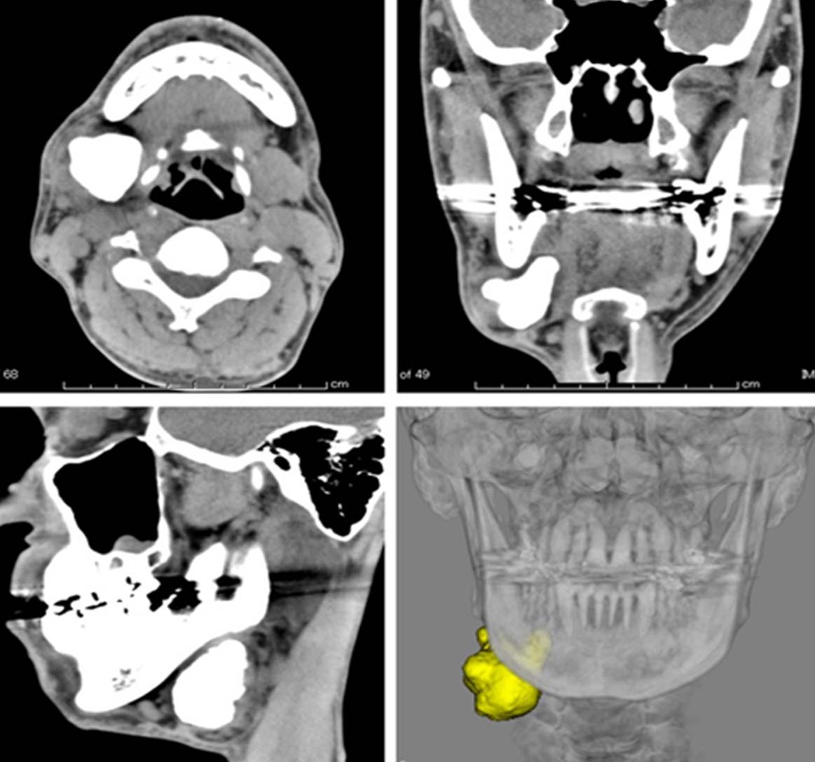
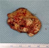
Evaluation of the bone window on CT revealed a lesion 41.2 mm × 32.1 mm × 27.1 mm in size. Three-dimensional evaluation using Mimics software (Materialise, Leuven, Belgium) gave a volume of 14.9 cm3. The clinical and radiological diagnosis was a giant sialolith in the parenchyma of the submandibular gland. No neurological disturbance of the facial, lingual, or sublingual nerve was evident. The patient underwent submandibular sialoadenectomy under general anaesthesia. A submandibular skin and platysma muscle incision was made and the flap was elevated. Careful dissection of the soft tissue was performed and the capsule of the submandibular gland was exposed. After careful dissection around the submandibular gland, the giant sialolith was removed first through the submandibular gland incision in order to make space for the approach to the lingual side of the submandibular gland. The sialolith measured 41mm × 31mm × 26 mm [Table/Fig-2].
A giant sialolith of the submandibular gland.
Wharton’s duct was then ligated and severed and the submandibular ganglion resected, with preservation of the lingual nerve. There were no postoperative complications such as disturbance of the facial or lingual nerve. The postoperative course was uneventful.
Discussion
Sialolithiasis is one of the most common diseases of the salivary gland. Sialoliths tend to develop in the submandibular gland and can vary in size from less than a millimeter to a few centimeters in largest diameter, but are typically less than 10 mm in diameter [1]. Giant sialoliths are defined as those exceeding 15 mm in any one dimension [1] and are commonly located in Wharton’s duct [1,2]. Sialoliths are thought to enlarge at a rate of approximately 1 mm to 1.5 mm per year [3], and in our case, the sialolith had been growing for about 40 years, resulting in a size of about 40 mm.
In a survey of 2,959 sialoliths by Sigismund PE et al., lesion size ranged from 10 mm to 35 mm. Giant sialoliths (>15 mm) are rare and those in Wharton’s duct are commonly cylindrical or elongated [4]. In contrast, intraparenchymal giant sialoliths are ovoid or irregular. Ledesma-Montes C et al., reviewed 16 cases of giant sialoliths measuring ≥35 mm, all of which were in male patients aged >34 years [2]. The sialoliths varied from 35 mm to 70 mm; only one sialolith was located in Stensen’s duct of the parotid gland, and five of the remaining 15 cases (33.3%) of submandibular gland sialoliths were located in the parenchyma. The parenchymal sialoliths were between 35 mm and 55 mm in length. To our knowledge, the largest sialolith reported in the literature to date was 83 mm in length in Wharton’s duct [5].
Bodner evaluated imaging modalities for the diagnosis of giant sialoliths and found panoramic radiograph and axial CT were comparable in the precise preoperative estimation of stone size [1]. In the present case, the size of the actual sialolith was almost the same as that measured on CT (bone window). However, Arslan S et al., reported a giant submandibular sialolith of 42 mm × 17 mm in size on axial CT and of 35 mm × 25 mms × 15 mm in actual size [6]. Because the measurement on CT was not performed using the bone window setting, the size on CT was larger than the actual size. In the second largest sialolith of Wharton’s duct reported, which was 72 mm in length when the fragments of sialolith were realigned after removal [5], the length was underestimated from a panoramic radiograph at approximately 65 mm. Therefore, linear measurement of the specimen may not be an accurate indication of stone size. To evaluate the size of sialoliths accurately, we propose CT (bone window)-based linear and volume measurement of giant sialoliths using software.
[1]. Bodner L, Giant salivary gland calculi: diagnostic imaging and surgical managementOral Surg Oral Med Oral Pathol Oral Radiol Endod 2002 94:320-23. [Google Scholar]
[2]. Ledesma-Montes C, Garcӑs-Ortíz M, Salcido-García JF, Hernández-Flores F, Hernández-Guerrero JC, Giant sialolith: case report and review of the literatureJ Oral Maxillofac Surg 2007 65:128-30. [Google Scholar]
[3]. Iqbal A, Gupta AK, Natu SS, Gupta AK, Unusually large sialolith of Wharton’s ductAnn Maxillofac Surg 2012 2:70-73. [Google Scholar]
[4]. Sigismund PE, Zenk J, Koch M, Schapher M, Rudes M, Iro H, Nearly 3,000 salivary stones: some clinical and epidemiologic aspectsLaryngoscope 2015 125:1879-82. [Google Scholar]
[5]. Shahoon H, Farhadi S, Hamedi R, Giant sialoliths of Wharton duct: Report of two rare cases and review of literatureDent Res J (Isfahan) 2015 12:494-97. [Google Scholar]
[6]. Arslan S, Vuralkan E, Çobanog˘lu B, Arslan A, Ural A, Giant sialolith of submandibular gland: report of a caseJ Surg Case Rep 2015 4:rjv043 [Google Scholar]