Primary Endometrial Squamous Cell Carcinoma in-situ with Extensive Icthyosis Uteri: A Rare Case Report
Manjula Jain1, Anamika Kashyap2, Ratna Biswas3
1 Director Professor, Department of Pathology, Lady Hardinge Medical College, New Delhi, India.
2 Senior Resident, Department of Pathology, Lady Hardinge Medical College, New Delhi, India.
3 Director Professor, Department of Obstetrics and Gynecology, Lady Hardinge Medical College, New Delhi, India.
NAME, ADDRESS, E-MAIL ID OF THE CORRESPONDING AUTHOR: Dr. Anamika Kashyap, Senior Resident, Department of Pathology, Lady Hardinge Medical College, New Delhi-110001, India.
E-mail: anamikakashyap25@gmail.com
Primary squamous cell carcinoma of the endometrium is a rare entity with primary endometrial squamous cell carcinoma in-situ being more uncommon. We report a 60-year-old multiparous post-menopausal woman who presented with a lower abdominal swelling alongwith difficulty in urination for five months. Total abdominal hysterectomy with bilateral salpingo-oophorectomy showed an enlarged uterus with pyometra. A diagnosis of primary squamous cell carcinoma in-situ of the endometrium was made on histopathology.
Endometrium, Pyometra, Uterus
Case Report
A 60-year-old multiparous post-menopausal woman presented with lower abdominal swelling and urinary discomfort for five months. There was no history of vaginal bleeding and the patient had been post-menopausal for 15 years. Abdominal examination revealed a soft, nontender lump corresponding to the size of 24 weeks pregnant uterus. A speculum examination showed cervical stenosis. Pelvic ultrasound showed an enlarged uterus with accumulation of around one litre fluid within the endometrial cavity. A Papanicolaou smear of the cervix was negative for intraepithelial lesions or malignancy. Total abdominal hysterectomy with bilateral salpingo-oophorectomy was performed. One litre of pus was drained from uterus per operatively.
On gross examination, the uterus was enlarged with a thinned out cervix with focal areas of ulceration. There was no evidence of any cervical growth. Endometrial cavity was distended with an irregular, shaggy, grey yellow endometrial surface. Myometrium and bilateral adnexae were unremarkable [Table/Fig-1a]. Almost entire endometrium was replaced by stratified squamous epithelium with extensive areas of moderate dysplasia and carcinoma in-situ with presence of occasional atrophic endometrial glands covered by areas of ulceration [Table/Fig-1b,c].
a) Hysterectomy specimen irregular, shaggy, grey yellow endometrial surface; b) Extensive replacement of endometrial lining by sheets of squamous cells with dysplasia on left (H&E 10X); c) Endometrial squamous cell carcinoma in situ with occasional atrophic endometrial gland (H&E 10X); d) Immunohistochemistry showing high Ki-67 labelling index (IHC 10X).
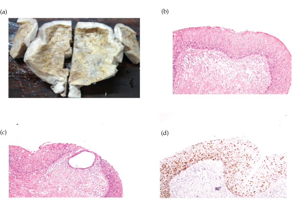
Underlying myometrium was unremarkable. Cervix showed extensive areas of ulceration with focal squamous metaplasia. However, no evidence of dysplasia was seen on processing the entire cervix. Also, there was no evidence of malignant endometrial gland on extensive sectioning. Based on these histopathology findings and a high Ki- 67 labelling index [Table/Fig-1d] on immunohistochemistry, a final diagnosis of icthyosis uteri with extensive dysplasia and foci of primary endometrial squamous cell carcinoma in situ-was given. Patient is disease free, three months after surgery with negative Papanicolaou smear of vaginal vault.
Discussion
Primary Endometrial Squamous Cell Carcinomas (PESCC) have been found in association with pyometra, cervical stenosis, chronic inflammation, multiparity and icthyosis uteri in post-menopausal women. An icthyosis uterus is a rare condition of replacement of the entire endometrial surface by stratified squamous epithelium [1]. PESCC can arise from endometrial stem cell, squamous metaplasia of the normal endometrium or from heterotopic cervical tissue [2]. The presence of squamous epithelium in the endometrium is termed ichthyosis uteri, leukoplakia, epidermalization psoriasis uteri, epidermoid heteroplasia, cholesterometra, acanthosis and indirect regenerative squamous metaplasia [2].
Chronic irritation due to long standing pyometra may lead to the malignant change in icthyosis uteri as seen in the present case. Primary endometrial squamous cell carcinoma in-situ arising in ichthyosis uteri is a very rare entity with a very few case reports in the literature. To the best of our knowledge, only six cases of PESCC in situ have been reported to date [3-8]. Pailoor K et al., and Jetley S et al., had described similar rare case of PESCC in-situ with extensive ichthyosis uteri in a post-menopausal woman with pyometra [3,4].
To reach a diagnosis of PESCC, it is important to exclude cervical SCC extension into the endometrium and squamous differentiation of an endometrioid adenocarcinoma. PESCC can be differentiated from endometrial SCC involvement by pathological criteria recommended by Fluhmann: (1) no evidence of a co-existing endometrial adenocarcinoma or primary cervical SCC; (2) no connection between the endometrial tumour and squamous epithelium of the cervix; (3) no connection between any in-situ carcinoma of cervix and endometrial neoplasm [9]. Our present case fulfilled Fluhmann’s criteria with presence of changes of dysplasia and carcinoma in-situ without any signs of an invasive carcinoma even on extensive sampling.
A high Ki-67 labelling index has also been reported, explaining the aggressive and malignant nature of the lesion. The prognosis for patients with endometrial SCC depends on the stage of the tumour. PESCC shows a poorer prognosis than endometrioid carcinomas and is managed by surgical hysterectomy with salpingo-oopherectomy and radiotherapy [9].
Conclusion
Primary squamous cell carcinoma of endometrium although rare, should be considered as a diagnosis while evaluating a post-menopausal elderly females presenting with pyometra. Early prompt diagnosis is important to improve the survival rate.
[1]. Bagga PK, Jaswal TS, Datta U, Mahajan NC, Primary endometrial squamous cell carcinoma with extensive squamous metaplasia and dysplasiaIndian J Pathol Microbiol 2008 51(2):267-68. [Google Scholar]
[2]. Lee SJ, Choi HJ, Primary endometrial squamous cell carcinoma: A case report and review of relevant literature on Korean womenKorean J Pathol 2012 46:395-98. [Google Scholar]
[3]. Pailoor K, Pai MR, Gatty RC, Fernandes H, Jayaprakash CS, Marla NJ, A rare case of primary insitu squamous cell carcinoma of the endometrium with extensive icthyosis uteriOnline J Health Allied Sci 2014 13:10 [Google Scholar]
[4]. Jetley S, Jairajpuri ZS, Hassan MJ, Madaan G, Jain R, Primary endometrial squamous cell carcinoma in situ report of a rare diseaseSQU Medical Journal 2015 15(4):559-62. [Google Scholar]
[5]. Radhika S, Dey P, Gupta SK, Primary squamous cell carcinoma in-situ of the endometrium: A case reportIndian J Cancer 1993 30:92-95. [Google Scholar]
[6]. Mitchell S, Fletcher H, Williams NP, Coard K, In-situ squamous cell carcinoma of the endometrium associated with long-term intrauterine device (Dalkon Shield) usageJ Obstet Gynaecol 1999 19:88-89. [Google Scholar]
[7]. Zidi YS, Bouraoui S, Atallah K, Kchir N, Haouet S, Primary in-situ squamous cell carcinoma of the endometrium, with extensive squamous metaplasia and dysplasiaGynecol Oncol 2003 88:444-46. [Google Scholar]
[8]. Kairys LR, Dougherty CM, Mickal A, Squamous cell carcinoma in-situ of the endometrial cavityAm J Obstet Gynaecol 1964 88:548-89. [Google Scholar]
[9]. Fluhmann CF, The histogenesis of squamous cell metaplasia of the cervix and endometriumSurg Gynecol Obstet 1953 97:45-58. [Google Scholar]