Introduction
During the last 200 years, there have been many changes in the way of performing endodontic treatment. The standard protocol has undergone several modifications, more so because of increased demand from the patients for saving their teeth and advances in material science and innovative equipments. Bioceramics materials in endodontics can be considered as a magnanimous entity which has changed the prognosis of many cases which were once considered as next to impossible.
A remarkable biocompatible material, MTA with exciting clinical applications was pioneered by Dr. Mahmoud Torabinejad and co-workers in Loma Linda University [1]. MTA can be used in surgical and non–surgical applications, including direct pulp capping [2], temporary filling material, Perforation repairs in roots or furcations [3], apexification and root end fillings [4,5]. Despite the high clinical efficacy of this wonder cement, there were always some issues which prevented the clinicians to use it for many cases. The major ones being very long setting time and difficult manipulation.
Biodentin new bioactive calcium silicate-based cement has been recently launched in the dental market as a ‘dentin substitute’. This new biologically active material aids its penetration through opened dentinal tubules to crystallize interlocking with dentin and provide mechanical properties. Biodentin has been formulated using MTA-based cement technology and hence; claims improvements of some of the properties such as physical qualities and handling, including its other wide range of applications like endodontic repair and pulp capping in restorative dentistry [6].
This review article attempts to compile and compare the properties of MTA and Biodentin for better clinical understanding.
Chemical Composition
Chemical composition of Biodentine: Biodentine is available in the form of a capsule containing the ideal ratio of its powder and liquid. The composition of powder is given in [Table/Fig-1] while the liquid contains calcium chloride which act as an acclerator, hydrosoluble polymer function as water reducing agent and water. However, the exact concentration of its components has not been provided by the manufacturer, various researchers have studied the same and provided the data. One such study performed by Camilleri J et al., has revealed the concentration of components of Biodentine [Table/Fig-1] [7].
Composition of Biodentine.
| Powder | Percentage |
|---|
| Tricalcium silicate (3CaO.SiO2) (main core material)Dicalcium silicate (2CaO.SiO2) (second core material)Calcium carbonate (CaCO2)(filler)Zirconium Oxide (ZrO2) (radioopacifier)Iron oxide(colouring agent) | 80.1-14.95- |
Chemical composition of MTA: MTA is basically a mechanical mixture of three powder ingredients: portland cement (75%), bismuth oxide (20%) and gypsum (5%) [8]. According to MTA patent, it consist of calcium oxide (50-75 wt %) and silicon oxide (15-20 wt %), which together constitute 70-95% of the cement. Upon blending of these raw materials; tricalcium silicate, dicalcium silicate, tricalcium aluminate, tetracalcium aluminoferrite are produced [9]. There are two commercial types of MTA: grey and white and the difference lies due to the presence of iron in the former which further forms the tetracalciumalumino-ferrite phase. On the contrary, there is absence of oxide of iron in white MTA and hence the phase [Table/Fig-2] [10].
| Composition | Percentage |
|---|
| PowderTricalcium silicateDicalcium silicateTricalcium aluminateTetracalciumaluminoferrite Calcium sulphate Bismuth oxide Calcium oxide Silicon oxide Aluminium oxide | 66.18.42.0--1480.51.0 |
Setting Reaction
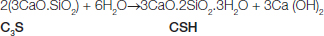
Setting reaction of Biodentine: The reaction of the powder with the liquid leads to the setting and hardening of the cement. Just after mixing, the calcium silicate particles of Biodentine react with water to from a high pH solution containing Ca2+, OH- and silicate ions. The hydration of the tricalcium silicate leads to the formation of a hydrated calcium silicate gel on the cement particles and calcium hydroxide nucleates. With the passage of time, calcium silicate hydrated gel polymerizes to form a solid network and the alkalinity of the surrounding medium increases due to the release of calcium hydroxide ions. Further the hydrated calcium silicate gel surrounds the unreacted tricalcium silicate particles and due to its relatively impermeable nature to water, it helps in slow down the effects of further reactions [11,12]. The complete reaction can be summarized as [6]:
Setting reaction of MTA: The hydration reaction during setting occurs between tricalcium silicate and dicalcium silicate to form a calcium hydroxide and calcium silicate hydrate gel, producing an alkaline pH. A further reaction between tricalcium aluminate and calcium phosphate forms a high-sulphate calcium sulphoaluminate. The calcium ions leach through the dentinal tubules, and the concentration increases with time as the material cures [8,13,14].
Setting Time
The working time of Biodentine and MTA is given in [Table/Fig-3] [6]. The presence of setting accelerator in Biodentine results in faster setting thereby improving its handling properties and strength. This is an advantage over MTA, since a delayed setting time studied by Torabinejad M et al, leads to an increased risk of partial material loss and alteration of the interface during the finishing phase of the procedure [5,13,14]. Therefore, Biodentine has a great improvement compared to MTA in terms of setting time.
Setting time and porosity characteristicsof MTA and Biodentine.
| Material | Initial setting time (minutes) | Final setting time (minutes) | Porosity characteristics {density(g/cm3)} |
|---|
| MTA (ProRoot) | 70 | 175 | 1.882 (0.002) |
| Biodentine | 6 | 10.1 | 2.260 (0.002) |
Density and Porosity
It is the critical factor which determines the amount of leakage and outcome of the treatment because greater pore diameter results in larger leakage which corresponds to the ingress and transmission of microorganisms and hence compromised hermetic seal. A study performed by Torabinejad M et al., [5] did not reveal any significant solubility of MTA whereas; Fridland M and Rosado R have reported the significant increase in solubility and porosity of ProRoot MTA with the increase in water to powder ratio [15]. De Souza ET et al., conducted a study on porosity and compared Biodentine with IRoot BP, Ceramicrete and ProRoot MTA using micro-CT characterization. They observed that no significant differences were found in porosity between the new calcium silicate containing repair cements and MTA [16].
Due to low water content in the mixing stage Biodentine exhibits lower porosity than MTA [Table/Fig-3].
Compressive Strength
During the setting of Biodentine, the compressive strength increases 100 MPa in the first hour and 200 MPa at 24th hour and it continues to improve with time over several days until reaching 300 MPa after one month [8] which is comparable to the compressive strength of natural dentine i.e 297 MPa [17]. A study conducted by Grech L et al., showed that Biodentine had highest compressive strength when compared to other tested materials due to low water/cement ratio used in Biodentine [14].
Flexural Strength
Flexural strength of any dental material is an important factor as it decreases the risk of fracture in clinical use. Walker MP et al., found that the flexural strength of MTA was 14.27 MPa when specimens were exposed to two-sided moisture after 24th hour of setting time [18]. However, the flexural strength of Biodentine recorded after two hours, has been found to be 34 MPa [6].
Microhardness
Microhardness of MTA has been affected by several factors like the pH value of the environment, the thickness of the material, the condensation pressure, the amount of entrapped air in the mixture, humidity, acid etching of the material, and temperature [19-22]. Matt GD et al., investigated the microhardness of grey MTA and white MTA with 2 mm and 5 mm thickness of material and found that 5 mm thickness has significantly more hardness regardless of the material used [19]. Grech L et al studied that Biodentine showed superior value of microhardness when compared to Bioaggregate and IRM [14]. The hardness of biodentine by Goldberg et al., was found to be 51 Vickers Hardness Number (VHN) at 2 hour and 69 VHN after one month [23]. The crystallization of calcium silicate hydrate gel continues which reduces porosity and increase hardness with time [6].
Radiopacity
The mean radiopacity for MTA has been found to be 7.17 mm of equivalent thickness of aluminium [5] and Biodentine reported a radiopacity to 3.5 mm of aluminium [6]. Grech L et al., evaluated the radiopacity of Biodentine, bioaggreagte and tricalcium silicate cement and found that all materials had radiopacity value greater than 3 mm of aluminium [14]. However Caron G et al., observed lower radiopacity of biodentine when compared to MTA Angelus [24].
Solubility
From the literature available, there is no definite conclusion regarding the degree of MTA solubility and it was concluded that with increase in water-to-powder ratio, release of calcium from MTA increases which accelerates its solubility [5,20,15]. Grech L et al., investigated lowest degree of solubility for Biodentine, bioaggregate and prototype cement while assessing the physical properties of these materials. They confirmed the deposition of hydroxyapatite crystals on material surface in presence of synthetic tissue fluid [14].
Microleakage
Biodentine is found to be associated with high pH (12) and releases calcium and silicon ions which stimulates mineralization and create “mineral infiltration zone” along dentin-cement interface imparting a better seal. Caron G et al., have found that Biodentine exhibits superior sealing properties than MTA [24]. While Torabinejad M reviewed a comprehensive literature to investigate studies regarding the leakage of MTA and concluded that MTA has good sealing ability and it seals well [25]. Ravichandra PV et al., evaluated that Biodentine provide better adaptation and seal than commonly used root-end filling material [26]. However, Ozbay G et al., observed less microleakage with MTA then Biodentine when analysed by fluid filteration method [11].
Marginal Adaptation and Sealing Ability
Marginal adaptation has correlation with the sealing ability of dental material and, hence effect on clinical success rate. Micromechanical adhesion of Biodentine enabled excellent adaptability of Biodentine crystals to the underlying dentin [27].
Soundappan S et al., conducted invitro study to compare the marginal adaptation of Biodentine with MTA and Intermediate Restorative Material (IRM) using scanning electrion microscope and concluded that both MTA and IRM were significantly superior to Biodentine in terms of marginal adaptation when used as a root end filling material [28].
Bond Strength and Push Out Bond Strength
Tunc ES et al., reported that the application of total-etch 1-bond adhesive system with a composite and compomer over MTA results significantly higher bond strength than with a 1-step self-etch adhesive system [29]. Whereas, Hashem DF et al., investigated that Biodentine has low strength during initial stages of setting, hence the application of final overlying resin composite restoration (laminated or layered) should be delayed for more than two weeks in order to achieve adequate bond strength of matured biodentine to withstand contraction forces of composite [30].
Another important use of MTA and Biodentine is perforation repair and to deliver higher success rate, the material should possess high push out bond strength which aids to prevent the dislodgement of material during tooth function from the repair site. El-Maaita AM et al., examined the effect of smear layer on push out bond strength of biodentine, ProRoot MTA and Harvard MTA and reported that push out bond strength decreases with removal of smear layer in all tested materials [31]. Guneser MB et al., assessed the effect of different endodontic irrigating agents (sodium hypochlorite, chlorhexidine and saline) on push out bond strength of MTA and Biodentine when used as a root perforation repair material and concluded that MTA possess low push out bond strength than Biodentine [32].
Discoloration
Literature reveals that presence of transitional elements namely iron, manganese, copper and chromium impart strong color to the material in it oxide forms. In the same way, bismuth, heavier element causes discoloration owing to its yellow oxide.
Valles M et al., conducted an in vitro study to evaluate color stability of five calcium silicate based material under the influence of light and oxygen and found that combination of light and anaerobic conditions results in significant differences in color of Angelus white MTA, ProMTA, white portland cement with bismuth oxide whereas Biodentine and PC exhibited color stability for five days [33].
Antibacterial and Antifungal Activity
The antibacterial and antifungal properties of MTA and Biodentine can be best attributed to the high pH of these materials. This high alkalinity has inhibitory effect on the growth of microorganism and causes disinfection of dentin. Hiremath GS et al., evaluated the antimicrobial efficacy of Biodentine, MTA, and MTA Plus and found that MTA and Biodentine showed significant antimicrobial effect against E. faecalis whereas MTA Plus was proved to be a good anti fungal agent against Candida albicans [34]. Another study reported the antibacterial and anti fungal characteristics of Biodentine, MTA and Glass Ionomer cement (GIC) and concluded that Biodentine shows superior antimicrobial action than MTA and GIC [35].
Biocompatibility and Cytotoxicity
Biocompatibility and cytotoxicity of a dental material have to be taken care of when material is used as a perforation or furcal repair, retrograde filling or pulp capping agent to avoid its toxic effect on surrounding tissue (pulpal and periradicular cells). Zhou H et al., compared the cytotoxicity of biodentine with white MTA and GIC using human gingival fibroblast and observed that biodentine caused similar reaction as compared to white MTA, and both materials were less cytotoxic than GIC [36]. Similarly, Nunez CMC et al., have found similar pattern of cytokine expression between Biodentine and MTA while using fibroblast cell [37]. Perard M et al., compared biocompatibility and gene expression of Biodentine and MTA using three-dimensional multicellular spheroid cultures and observed similar response between these two materials indicating its use for direct pulp-capping [38].
Bioactivity and Regenerative Potential
Laurent P et al., assessed the ability of Biodentine, MTA, calcium hydroxide and Xeno III adhesive resin to induce reparative dentin synthesis and transforming growth factor beta 1 (TGF-b1) secretions. They showed that both bBodentine and MTA involved in early odontoblastic differentiation and initiation of mineralisation and thus form reparative dentin synthesis then other two materials. TGF-b1 secretion was significantly increased with Biodentine, MTA and csalcium hydroxide than Xeno III [39].
In another study, Biodentine showed greater ability to produce apatite crystals and release of dental elements than MTA and BC sealer [40]. Luo Z et al., studied the effect of Biodentine on the human dental pulp stem cells (hDPSCs) and found that biodentine significantly increased the proliferative, migratory and adhesion of stem cells when placed directly in contact with the pulp, which further reflects bioactivity and biocompatibility properties of the material. Biodentine is able to promote mineralisation, generating a reactionary dentine as well as a dense dentine bridge when place in contact with pulp [41].
Bonson S et al., observed differentiation of fibroblasts and bone formation when MTA was placed on cell cultures of gingival and periodontal ligament fibroblasts. Thus, MTA is considered as a bioactive material with osteoinductive properties [42].
Clinical Implications and Limitations of MTA and Biodentine
The clinical applications and limitations/drawbacks of MTA and Biodentine are summarized in a tabular form [Table/Fig-4,5 and 6].
| Clinical applications | Related studies |
|---|
| Pulp capping | Primary teeth: Tuna D and Olmez A 2008 [43]Permanent teeth: Witherspoon DE [44] concluded that MTA is a good substitute of Ca(OH)2 for vital pulp therapies because it stimulates a higher and greater quality and quantity of reparative dentin and also aids superior long term sealing ability |
| Pulpotomy | Primary teeth: Torabinajad M and Chivin N [4], Ni Chaollai A et al., 2009 [45]Permanent teeth: Belobrov I et al., 2008 [46], Eghbal MJ et al., [47] revealed that MTA pulpotomy forms a dentine bridge completely and maintains the vitality of the radicular pulp by limiting the inflammation. |
| Perforation repair (Furcal or root) | Primary teeth: Oliveria TM et al., 2008 [48]Permanent teeth: Arens DE and Torabinajad M [3], Wang P et al., 2009 [49] (MTA promotes bone healing and eliminates the signs of inflammation. hence, MTA can be considered as an alternative option for repair of furcal perforations both in primary and permanent teeth). |
| Root end filling | Permanent teeth: Christiansen R et al., 2009 [50] found that healing of teeth treated with MTA as root end filling material had significantly better healing (96%) than those treated with orthograde GP filling (52%). |
| Root canal filling | Primary teeth (absence of permanent successor): O’SullivanSM and Hartwell GR 2001 [51] Permanent teeth: Bogen G and Kuttler S 2009 [52] (obturation with MTA seemed to provide a biocompatible seal of root canal system). |
| Resorption | Primary teeth: Sari S and Sonmez D 2009 [53] Permanent teeth: Silveria FF et al., [54] treated a double “Pink tooth” with MTA as root canal filling material and found favourable results after 18 months. Hence, MTA may be suitable material for treatment of internal resorption. |
Clinical uses of Biodentine.
| Clinical applications | Related studies |
|---|
| Pulp capping | Laurent P et al., (39), Nowicka A et al., [55] (When Biodentine was applied directly onto pulp, it induces a reparative dentine formation due to modulation of pulp cell TGF-Beta secretion). |
| Pulpotomy | Villat C et al., [56] demonstrated that Biodentine should be considered as a conservative intervention in treatment of symptomatic immature teeth. |
| Root end filling | Pawar AM et al., 2013 [57] showed that routine endodontic therapy followed by surgical intervention with placement of biocompatible material like Biodentine for management of chronic peripapical lesions would positively affect the treatment outcome. |
| Dentin substitute | Koubi G et al., [58] conducted a randomized 3 years prospective study and concluded that Biodentine can be used as a dentine substitute under composite for posterior restorations. |
| Perforation repair (Furcal or root) | Hassan FN et al., [59] found that both ProRoot MTA and Biodentine performed equally well when used as furcation repair materials. |
| Root canal filling and apex closure | Han L and Okiji T 2013 [40], Elumalai D et al., 2015 [60] described that that initial healing was better in case of Biodentine when using in open apices cases however MTA showed better long term effects. |
Limitations of MTA and Biodentine.
| Drawbacks | Related Studies |
|---|
| MTA Long setting time | Torabinajad M et al., 1995 [5], Chng HK et al., 2005 [61] (MTA showed longest setting time when compared to that amalgam). |
| Difficult handling | Mooney GC and North S 2008 [62] observed that the manipulation of MTA was messy when the moisture was excessive in the preparation which further results in soupy material and hence difficult to use |
| Discoloration | Accorinte ML et al., 2008 [63] (iron and manganese were the possible elements responsible for discoloration). |
| Toxic elements in composition | Asgary S et al., 2006 [64] (MTA contains elements like arsenic which diffused into the tissue fluids and could potentially cause toxicity). |
| High cost |
| BIODENTINEPoor radio-opacity | Caron G et al., 2014 [24] (despite the presence of zirconium dioxide, Biodentine has unfavourable radiopacity as compare to MTA). |
| Lower wash out resistance | Grech L et al., 2013 [14], Elumalai D et al., 2015 [60] demonstrated that Biodentine has a high washout, low fluid uptake and sorption values, low setting time and superior mechanical properties. |
Conclusion
The clinical uses of bioceramics have increased exponentially over the years because of their wide range of applicability in restorative dentistry and endodontics. The introduction of MTA was considered as a major break-through in the history of material science and since then the properties of this material have been improvised in order to achieve its maximum benefits. However, there have been a few limitations of this material which have always compelled the researchers worldwide to look for its alternatives. Difficult manipulation, slow setting time and high cost are the ones to name a few. In order to overcome these limitations, a new bioceramic material named Biodentine was introduced in the year of 2010 which has proved to be a second major break-through. Relatively easier manipulation, low cost and faster setting is the major advantages of this material when compared to MTA. Studies have also proved that its compressive and flexural strength are superior to that of MTA. High biocompatibility and excellent bioactivity further go in favour of this dental replacement material. Due to lack of long term observational studies, it is difficult to infer concretely that which material out of MTA and Biodentine is superior, however, manoeuvrability and economical factors fall in favour of Biodentine.
[1]. Torabinejad M, Watson TF, Pitt Ford TR, Sealing ability of a mineral trioxide aggregate when used as a root end filling materialJ Endod 1993 19(12):591-95. [Google Scholar]
[2]. Pitt Ford TR, Torabinejad M, Abedi HR, Bakland LK, Kariyawasam SP, Using mineral trioxide aggregate as a pulp capping materialJ Am Dent Assoc 1996 127(10):1491-94. [Google Scholar]
[3]. Arens DE, Torabinejad M, Repair of furcal perforations with mineral trioxide aggregate: two case reportsOral Surg Oral Med Oral Pathol Oral Radiol Endod 1996 82(1):84-88. [Google Scholar]
[4]. Torabinajad M, Chivian N, Clinical applications of mineral trioxide aggregateJ Endod 1999 25(3):197-205. [Google Scholar]
[5]. Torabinejad M, Hong CU, McDonald F, Pitt Ford TR, Physical and chemical properties of a new root-end filling materialJ Endod 1995 21(7):349-53. [Google Scholar]
[6]. Septodont Biodentine™ Active Biosilicate Technology™Scientific file 2010 [Google Scholar]
[7]. Camilleri J, Sorrentino F, Damidot D, Investigation of the hydration and bioactivity of radiopacified tricalcium silicate cement, Biodentine and MTA AngelusDental Materials 2013 29(5):580-93. [Google Scholar]
[8]. Sarkar NK, Caicedo R, Ritwik P, Moiseyeva R, Kawashima I, Physiochemical basis of the biologic properties of mineral trioxide aggregateJ Endod 2005 31(2):97-100. [Google Scholar]
[9]. Torabinejad M, White DJ, Tooth filling material and useUS Patent Number 5,769,638 1995 [Google Scholar]
[10]. Camilleri J, The chemical composition of mineral trioxide aggregateJ Conserv Dent 2008 11(4):141-43. [Google Scholar]
[11]. Ozbay G, Kitiki B, Peker S, Kargul B, Apical sealing ability of a novel material: analysis by fluig filtration techniqueActa Stomatol Croat 2014 48(2):132-39. [Google Scholar]
[12]. Allen AJ, Thomas JJ, Jennings HM, Composition and density of nanoscale calcium-silicate-hydrate in cementDent Mater 2007 6(4):311-16. [Google Scholar]
[13]. Parirokh M, Torabinejad M, Mineral trioxide aggregate: A comprehensive literature review- Part III: Clinical applications, drawbacks, and mechanism of actionJ Endod 2010 36(3):400-13. [Google Scholar]
[14]. Grech L, Mallia B, Camilleri J, Investigation of the physical properties of tricalcium silicate cement-based root-end filling materialsDent Mater 2013 29(2):20-28. [Google Scholar]
[15]. Fridland M, Rosado R, Mineral Trioxide Aggregate (MTA), solubility and porosity with different water-to-powder ratiosJ Endod 2003 29(12):814-87. [Google Scholar]
[16]. De Souza ET, Nunes Tameirao MD, Roter JN, De Assis JT, De Almeida Neves A, De-Deus GA, Tridimensional quantitative porosity characterization of three set calcium silicate based repair cement for endodontic useMicrosc Res Tech 2013 76(10):1093-98. [Google Scholar]
[17]. O’Brien W, Dental Materials and their Selection 2008 [Google Scholar]
[18]. Walker MP, Diliberto A, Lee C, Effect of setting conditions on mineral trioxide aggregate flexural strengthJ Endod 2006 32(4):334-36. [Google Scholar]
[19]. Matt GD, Thorpe JR, Strother JM, McClanahan SB, Comparative study of white andgray mineral trioxide aggregate (MTA) simulating a one- or two-step apical barriertechniqueJ Endod 2004 30(12):876-89. [Google Scholar]
[20]. Danesh G, Dammaschke T, Gerth HU, Zandbiglari T, Schafer E, A comparative study of selected properties of ProRoot mineral trioxide aggregate and two Portland cementsInt Endod J 2006 39(3):213-29. [Google Scholar]
[21]. Nekoofar MH, Adusei G, Sheykhrezae MS, Hayes SJ, Bryant ST, Dummer PM, The effect of condensation pressure on selected physical properties of mineral trioxideaggregateInt Endod J 2007 40(6):453-61. [Google Scholar]
[22]. Parirokh M, Torabinejad M, Mineral trioxide aggregate: A comprehensive literature review—Part I: Chemical, physical, and antibacterial propertiesJ Endod 2010 36(1):16-27. [Google Scholar]
[23]. Goldberg M, Pradelle-Plasse N, Tran XV, Colon P, Laurent P, Aubut V, Goldberg M, Emerging trends in (bio) material research Physico – chemical properties of BiodentineBiocompatibility or cytotoxic effects of dental composites 2009 1st edOxfordCoxmoor publishing co:181-203. [Google Scholar]
[24]. Caron G, Azerad J, Faure MO, Machtou P, Yves B, Use of a new retrograde filling material (Biodentine) for endodontic surgery: two case reportsInt J Oral Sci 2014 6(4):250-53. [Google Scholar]
[25]. Torabinejad M, Mineral trioxide aggregate: A comprehensive literature review—Part II: Leakage and BiocompatibilityJ Endod 2010 36(2):190-202. [Google Scholar]
[26]. Ravichandra PV, Vemisetty H, Deepthi K, Reddy J, Ramkiran D, Krishna JN, Comparative evaluation of marginal adaptation of biodentine and other commonly used root end filling materials - an in vitro studyJ Clin Diagn Res 2014 8(3):243-45. [Google Scholar]
[27]. Malkondu O, Kazandag MK, Kazazoglu E, A review on biodentine, a contemoprary dentin replacement and repair materialBioMed Research International 2014 :10Article ID 160951 [Google Scholar]
[28]. Soundappan S, Sundaramurthy JL, Raghu S, Natanasabapathy V, Biodentine versus mineral trioxide aggregate versus intermediate restorative material for retrograde root end filling: An in vitro studyJ Dent, Tehran Univ Med Sciences 2014 11(2):143-49. [Google Scholar]
[29]. Tunc¸ ES, Sonmez IS, Bayrak S, Egilmez T, The evaluation of bond strength ofa composite and a compomer to white mineral trioxide aggregate with twodifferent bonding systemsJ Endod 2008 34(5):603-65. [Google Scholar]
[30]. Hashem DF, Foxton R, Manoharan A, Watson TF, Banerjee A, “The physical characteristics of resin composite-calcium silicate interface as part of a layered/laminate adhesive restoration”Dent Mater 2014 30(3):343-39. [Google Scholar]
[31]. El-Maaita AM, Qualtrough AJE, Watts DC, “The effect of smear layer on the push-out bond strength of root canal calcium silicate cements”Dent Mater 2013 29:797-803. [Google Scholar]
[32]. Guneser MB, Akbulut MB, Eldeniz AU, “Effect of various endodntic irrigants on the push-out bond strength of biodentine and conventional root perforation repair materials”J Endod 2013 39(3):380-84. [Google Scholar]
[33]. Valles M, Mercade M, Duran-Sindren F, Bourdelande JL, Roig M, Influence of light and oxygen on the color stability of five calcium silicate-based materialsJ Endod 2013 39(4):525-28. [Google Scholar]
[34]. Hiremath GS, Kulkarni RD, Naik BD, Evaluation of minimal inhibitory concentration of two new materials using tube dilution method: An in vitro studyJ Conser Dent 2015 18(2):159-62. [Google Scholar]
[35]. Bhavna V, Chaitanya KP, Gandi P, Patel J, Dola B, Reddy RB, Evaluation of antibacterial and antifungal activity of new calcium-based cement (Biodentine) compared to MTA and glass ionomer cementJ Conser Dent 2015 18(1):44-46. [Google Scholar]
[36]. Zhou H, Shen Y, Wang Z, Li L, Zheng Y, Hakkinen L, In vitro cytotoxicity evaluation of a novel root repair materialJ Endod 2013 39(4):478-83. [Google Scholar]
[37]. Nunez CMC, Bosomworth MHJ, Field C, Whitworth JM, Valentine RA, Biodentine and mineral trioxide aggregate induce similar cellular responses in a fibroblast cell lineJ Endod 2014 40(3):406-11. [Google Scholar]
[38]. Perard M, Le Clerc J, Meary F, Perez F, Tricot-Doleux S, Pellen-Mussi P, Spheroid model study comparing the biocompatibility of Biodentine and MTAJ Mater Sci Mater Med 2013 24(6):1527-34. [Google Scholar]
[39]. Laurent P, Camps J, About I, Biodentine (TM) induces TGF-b1 release from human pulp cells and early dental pulp mineralizationInt Endod J 2012 45(5):439-48. [Google Scholar]
[40]. Han L, Okiji T, Bioactivity evaluation of three calcium silicate based endodontic materialsInt Endod J 2013 46(9):804-14. [Google Scholar]
[41]. Luo Z, Li D, Kohli MR, Yu Q, Kim S, He WX, Effect of Biodentine on the proliferation, migration and adhesion of human dental pulp stem cellsJ Dent 2014 42(4):490-97. [Google Scholar]
[42]. Bonson S, Jeansonne BG, Lallier TE, Root end filling materials alter fibroblasts differentiationJ Dent Res 2004 83(5):408-13. [Google Scholar]
[43]. Tuna D, Olmez A, Clinical long-term evaluation of MTA as a direct pulp capping material in primary teethInt Endod J 2008 41(4):273-78. [Google Scholar]
[44]. Witherspoon DE, Vital pulp therapy with new materials: new directions and treatment perspectives: permanent teethJ Endod 2008 34:25-28. [Google Scholar]
[45]. Ni Chaollai A, Monteiro J, Duggal MS, The teaching of management of the pulp in primary molars in Europe: a preliminary investigation in Ireland and the UKEur Arch Paediatr Dent 2009 10:98-103. [Google Scholar]
[46]. Belobrov I, Weis MV, Parashos P, Conservative treatment of a cervical horizontal root fracture and a complicated crown fracture: a case reportAust Dent J 2008 53(3):260-64. [Google Scholar]
[47]. Eghbal MJ, Asgary S, Baglue RA, Parirokh M, Ghoddusi J, MTA pulpotomy of human permanent molars with irreversible pulpitisAust Endod J 2009 35(1):04-08. [Google Scholar]
[48]. Oliveira TM, Sakai VT, Silva TC, Santos CF, Machado MA, Abdo RC, Repair of furcal perforation treated with mineral trioxide aggregate in a primary molar tooth:20-month follow-upJ Dent Child (Chic) 2008 75(2):188-91. [Google Scholar]
[49]. Wang P, Wang S, Ni L, The combination of a mineral trioxide aggregate and an adhesive restorative approach to treat a crown-root fracture coupled with lateral root perforation in a mandibular second molar: a case reportOper Dent 2009 34(4):497-502. [Google Scholar]
[50]. Christiansen R, Kirkevang LL, Hørsted-Bindslev P, Wenzel A, Randomized clinical trial of root-end resection followed by root-end filling with mineral trioxide aggregate or smoothing of the orthograde gutta-percha root filling: 1-year follow-upInt Endod J 2009 42(2):105-14. [Google Scholar]
[51]. O’Sullivan SM, Hartwell GR, Obturation of a retained primary mandibular second molar using mineral trioxide aggregate: a case reportJ Endod 2001 27(11):703-75. [Google Scholar]
[52]. Bogen G, Kuttler S, Mineral trioxide aggregate obturation: a review and case seriesJ Endod 2009 35(6):777-90. [Google Scholar]
[53]. Sari S, Sonmez D, Internal resorption treated with mineral trioxide aggregate in a primary molar tooth:18-month follow-upJ Endod 2006 32(1):69-71. [Google Scholar]
[54]. Silveira FF, Nunes E, Soares JA, Ferreira CL, Rotstein I, Double ‘pink tooth’ associated with extensive internal root resorption after orthodontic treatment: a case reportDent Traumatol 2009 25(3):e43-47. [Google Scholar]
[55]. Nowicka A, Lipski M, Parafiniuk M, Response of human dental pulp capped with biodentine and mineral trioxide aggregateJ Endod 2013 39(6):743-47. [Google Scholar]
[56]. Villat C, Grosgogeat B, Seux D, Farge P, Conservative approach of a symptomatic carious immature permanent tooth using a tricalcium silicate cement (Biodentine): A case reportRest Dent Endod 2013 38(4):258-62. [Google Scholar]
[57]. Pawar AM, Kokate SR, Shah RA, Management of a large periapical lesion using Biodentine as retrograde restoration with 18 months evident follow upJ Conser Dent 2013 6(6):573-75. [Google Scholar]
[58]. Kaubi G, Colon P, Franquin JC, Clinical evaluation of the performance and safety of a new dentine substitute, biodentine, in the restoration of posterior teeth-a prospective studyClin Oral Investig 2013 17(1):243-49. [Google Scholar]
[59]. Hassan FN, Al Hadi D, Saeed MM, Furcal perforation repair using MTA and biodentine: an in vitro evaluation using dye extraction methodInternational Journal of Recent Scientific Research 2015 6(3):3172-75. [Google Scholar]
[60]. Elumalai D, Kapoor B, Tewrai RK, Mishra SK, Comparision of mineral trioxide aggregate and biodentine for management of open apicesJournal of Interdesciplinary Dentistry 2015 5(3):131-35. [Google Scholar]
[61]. Chng HK, Islam I, Yap AU, Tong YW, Koh ET, Properties of a new root-end filling materialJ Endod 2005 31(9):665-68. [Google Scholar]
[62]. Mooney GC, North S, The current opinions and use of MTA for apical barrierformation of non-vital immature permanent incisors by consultants in paediatricdentistry in the UKDent Traumatol 2008 24(1):65-69. [Google Scholar]
[63]. Accorinte ML, Loguercio AD, Reis A, Carneiro E, Grande RH, Murata SS, Response of human dental pulp cappedwith MTA and calcium hydroxide powderOper Dent 2008 33(5):488-95. [Google Scholar]
[64]. Asgary S, Parirokh M, Eghbal MJ, Stowe S, Brink F, A qualitative X-ray analysis of white and grey mineral trioxide aggregate using compositional imagingJ Mater Sci Mater Med 2006 17(2):187-91. [Google Scholar]