According to Glossary of Periodontal Terminology (GPT), gingival recession is defined as location of gingival margin apical to Cemento-Enamel Junction (CEJ) [1]. The most common aetiological factor for the gingival recession and the loss of attached gingiva is abrasive and traumatic tooth brushing habits. The consequences of gingival recession are attachment loss, root exposure, which causes aesthetic concerns and root hypersensitivity, increases the risk of root caries and cervical abrasion [2]. Any surgical intervention for root coverage aims at restoring the marginal tissue to the CEJ and to regenerate the lost periodontium [3]. Many techniques have been tried for the treatment of gingival recession like pedicle grafts, autogenous free gingival grafts, subepithelial connective tissue grafts and combination of these techniques along with the techniques based on Guided Tissue Regeneration (GTR) principles [4-7]. Few limitations like second surgical site, technique sensitive, patient morbidity associated with procurement of autogenous free gingival grafts led to the usage of various other materials like Acellular Dermal Matrix (ADM), PRF membrane, AM and chorionic membrane [8-11]. Choukroun first developed the PRF in France [12]. It is a second generation platelet concentrate which is autologous and resorbable in nature entrapping various cytokines, growth factors and cells in its tetramolecular structure which are released over a period of time. Fibrin provides a matrix for migration of fibroblasts and endothelial cells, which are involved in the angiogenesis process and are responsible in the healing of new tissues [12]. Due to it’s certain disadvantages like requirement of blood withdrawal, expensive equipments and more time consumption during the procedures led the attention towards allografts like amnion membrane. AM is a thin, tough, transparent, avascular composite membrane composed of three major layers: a single epithelial layer, a thick basement membrane and an avascular mesenchyme made of collagen [13]. Presence of abundant laminin-5 helps in cellular adhesion of cells, growth of fibroblasts and neovascularisation in the early phases of wound healing. The matrix of human AM contains plenty of growth factor and has various biological properties such as anti-inflammatory, immunomodulatory, antimicrobial, antiviral and anti-scarring property. Owing to the presence of various growth factors, it induces angiogenesis, reduces the pain, and promotes epithelialization and extracellular matrix deposition [14]. However, there are only few studies comparing these two materials. Hence, the present study aims at evaluating and comparing the effectiveness of the PRF membrane and AM for the treatment of gingival recession by CAF technique.
Materials and Methods
This was an experimental design which was conducted on the patients visiting to the Department of Periodontics, Sri Aurobindo College of Dentistry, Indore, Madhya Pradesh, India with chief complain of unpleasant aesthetics and hypersensitivity. Systemically healthy subjects aged between 18-55 years with good oral hygiene maintenance after Phase-I therapy and Miller’s class I and II gingival recession were selected [15]. Patients were excluded from the study if they were poorly compliant, pregnant or lactating mothers, smokers, have undergone periodontal surgery in last six months or having fenestration and dehiscence. Overall, a maximum of 39 patients were deemed fit into inclusion-exclusion criteria. Out of 39 patients, nine patients dropped out from the study because they didn’t turn up for the surgical intervention after Phase-I therapy. The allocation of group to a patient was done randomly using simple random sampling technique by lottery method. Out of total 30 patients, 15 patients were allocated to one group treated as “Group A” (seven males and eight females) in which the root coverage was obtained by using platelet-rich fibrin membrane. The rest 15 patients (eight males and seven females) were allocated to second group treated as Group “B” in which the root coverage was obtained by using allograft AM. Ethical approval for the study was obtained from Ethical Committee of the Institute and prior consents for surgery from the patients were taken into account.
In Group- A and Group-B, root coverage was obtained by using coronally advanced flap technique along with placement of PRF membrane and dehydrated AM respectively. AMs of dimensions 1x1mm were obtained from Tata Memorial Hospital, Mumbai, India. Each group was followed up at three months and six months postoperatively. Acrylic stent was made and measurements were recorded at the deepest recession site with the pressure sensitive UNC-15 probe.
Clinical parameters which were measured: 1) PI [16]; 2) RD was measured from cemento-enamel junction to gingival margin by UNC-15 probe; 3) WKG was calculated with the UNC-15 probe by measuring the distance between gingival margin and mucogingival junction.
Surgical procedure: For all the selected patients, routine radiographic and blood investigations were done. Selected patients had little or no radiographic bone loss. Phase I therapy consisted of scaling, root planing, oral hygiene instructions and occlusal adjustments as needed. One week following Phase I therapy, a periodontal re-evaluation was performed.
Surgical technique: Both the groups were treated with CAF. Patients were instructed for prerinsing with 0.12% chlorhexidine solution for one minute after which scaling and root planing was carried out on exposed root surface. Horizontal incisions are given at the level of CEJ on the adjacent buccal papilla which are further joined by intrasulcular incision. Thereafter, two vertical incisions starting from the mesial and distal end of horizontal incision and extending beyond the mucogingival junction were given to raise a combination flap of full thickness flap coronally and partial thickness flap apically. De-epithelialization of papilla was done. After placement of selected membrane, the flap was coronally advanced and sutured. A tin foil and periodontal dressing was placed over the surgical area. All the patients were advised to use 0.2% chlorhexidine digluconate mouth rinse, twice daily. Systemic analgesics were prescribed and patients were advised to follow the routine postoperative instructions. The dressing and sutures were removed 10 days after surgery.
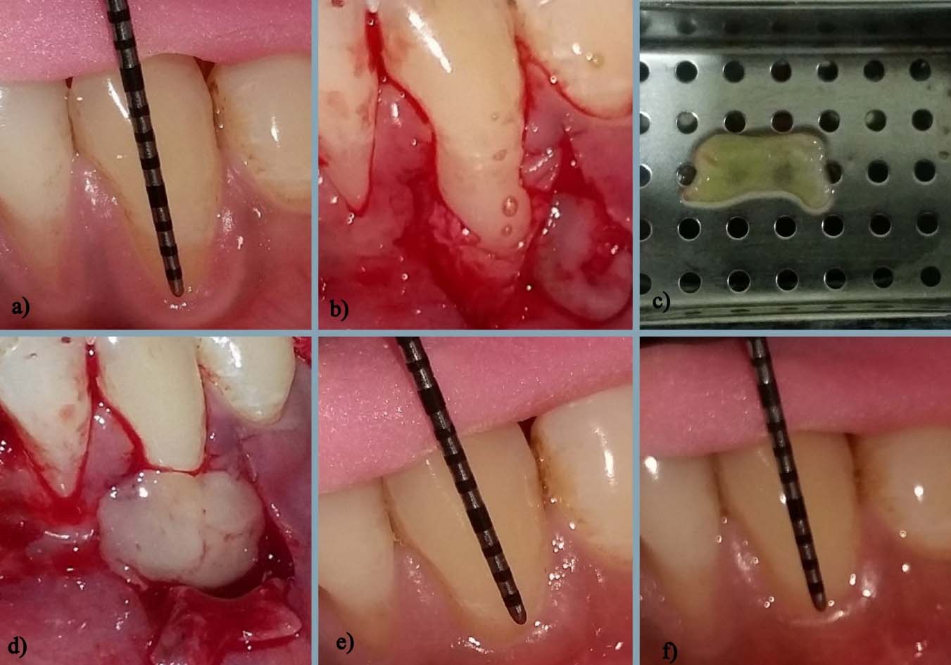
Group-A (PRF Group): The PRF was prepared following the protocol developed by Choukroun [12]. For Group A, 10 ml of intravenous blood (by a venipuncture of the antecubital vein) was withdrawn and collected in the centrifuge tubes without anticoagulant and was immediately centrifuged at 3000 revolutions/minute for 10 minutes. At the end of centrifugation, three layers were obtained, the top layer containing supernatant serum, the middle layer containing fibrin clot, and the bottom layer consisting of Red Blood Corpuscles (RBC). The fibrin clot was separated from the RBC base (preserving a small RBC layers) using sterile tweezers and scissors [12]. This fibrin clot was placed in PRF box for the preparation of the PRF membrane [Table/Fig-1].
a) Preoperative photograph; b) Flap reflection and recipient bed preparation; c) PRF membrane in PRF box; d) Placement of PRF membrane at surgical site; e) Three months postoperative; f) Six months postoperative.

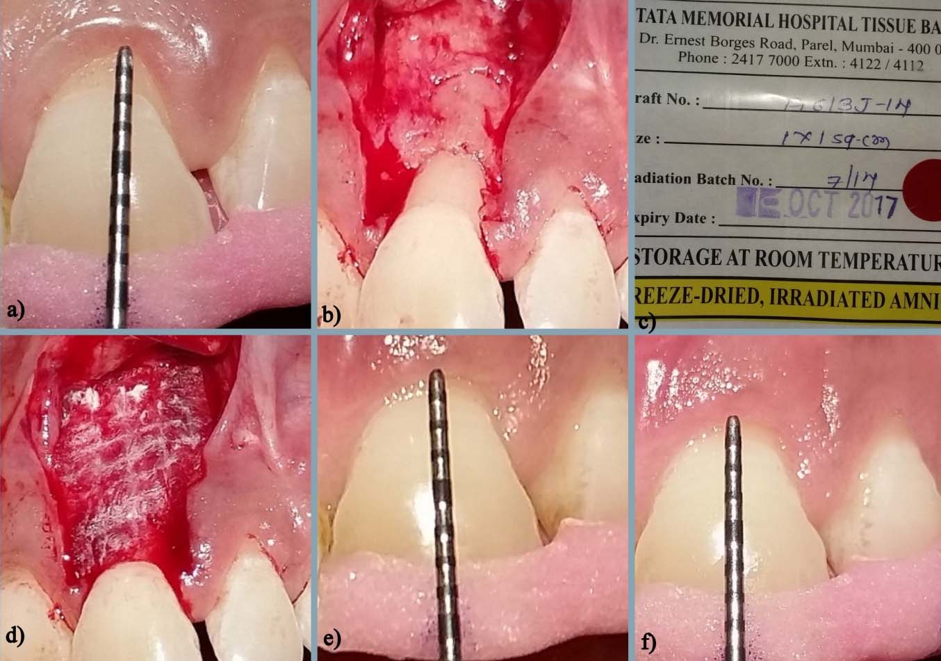
Group-B (Amnion Group): Same surgical technique as mentioned above was followed. The commercially available amniotic membrane (1x1 mm) was cut into the desired shape and length with scissors and placed onto the recession site [Table/Fig-2].
a) Baseline photograph; b) Flap reflection and recipient bed preparation; c) Commercially available amnion membrane; d) Amnion membrane placement; e) Three months postoperative; f) Six months postoperative.

Statistical Analysis
The raw data for 30 patients were entered into the computer database. Statistical software, SPSS version 17.0 trial was used for analysis. Incidence of an outcome variable along with 95% confidence limits was calculated. Mann-Whitney test was used to identify the significance of differences for all the parameters between Group-A and Group-B at three sampling stages (i.e., baseline, three months, and six months postoperative). Intragroup measurements between baseline and three months, three months and six months and baseline and six months, were carried out using the Wilcoxon signed ranks test. The probability value from p<0.05 to p<0.02 was considered as statistically significant while from p<0.01 to p<0.001 was considered as statistically highly/strongly significant.
Results
[Table/Fig-3] depicts statistically insignificant difference in the ages of the subjects of both the groups at the time of admission and thus age has not influenced the results. The mean age in Group-A and Group-B were found to be 27.27± 7.68 and 31.47±7.59 respectively.
The distribution of ages of patients included in Group A and Group-B.
| Variable | Scatter for age (year) | 95% Cl of the Mean Difference | t-value | LOS |
|---|
| Mean ± SD | LB | UB |
|---|
| Group A (Platelet rich fibrin) | 27.27±7.68 | 1.51 | 9.91 | 1.51 | p>0.05 * |
| Group B (Dehydrated amniotic membrane) | 31.47±7.59 |
| Mean difference | 4.20 year* |
The mean difference is not significant (insignificant) at the 0.05 level of significance. [Degrees of freedom is 28; UB-Upper Bound; LB-Lower Bound; LOS-Level of Significance] Mann-Whitney test was applied.
[Table/Fig-4] displays that there was no statistically significant differences in mean plaque index of patients of Group A based on scores between baseline and three months post surgery (p>0.05), at three months (p>0.05) and six months post surgery, and baseline and six months post surgery (p>0.05) as it couldn’t reach at the (p>0.05) level of significance. Similar findings were observed in the patients of Group-B.
Intragroup difference in PI between both the groups at baseline, three months and six months.
| Group | Sampling Stage | Plaque Index | Z-statistic ⇑ | LOS |
|---|
| Mean ± SD |
|---|
| Group A (Platelet rich fibrin) | Baseline | 0.860 | ±0.083 | 1.00 | p>0.05* |
| At 3 months | 0.840 | ±0.091 |
| At 3 months | 0.840 | ±0.091 | 1.67 | p>0.05* |
| At 6 months | 0.873 | ±0.080 |
| Baseline | 0.860 | ±0.083 | 0.63 | p>0.05* |
| At 6 months | 0.873 | ±0.080 |
| Group B (Amniotic membrane) | Baseline | 0.827 | ±0.122 | 1.73 | p>0.05* |
| At 3 months | 0.787 | ±0.106 |
| At 3 months | 0.787 | ±0.106 | 1.41 | p>0.05* |
| At 6 months | 0.813 | ±0.113 |
| Baseline | 0.827 | ±0.122 | 0.51 | p>0.05* |
| At 6 months | 0.813 | ±0.113 |
Wilcoxon signed ranks test.
The differences based on scores between groups are not significant (insignificant) at the 0.05 level of significance.
[Table/Fig-5] shows the results of intragroup comparison which suggests that there was significant reduction in recession depth between baseline and three months postoperative (p<0.001) and baseline and six months postoperative in both the groups (p<0.05) whereas statistically insignificant reduction was observed between the values of three months postoperative and six months postoperative (p>0.05) in both the groups individually.
Intra group comparison of the recession depth in both the groups A and B at baseline, three and six months postoperative.
| Group | Sampling stage | Recession depth (mm) | Z-statistic ⇑ | LOS |
|---|
| Mean ± SD |
|---|
| Group A (Platelet rich fibrin) | Baseline | 2.733 | ±0.799 | 3.25 | p<0.001§ |
| At 3 months | 1.333 | ±0.617 |
| At 3 months | 1.333 | ±0.617 | 1.00 | p>0.05* |
| At 6 months | 1.400 | ±0.633 |
| Baseline | 2.733 | ±0.799 | 3.13 | p<0.002§ |
| At 6 months | 1.400 | ±0.633 |
| Group B (Amniotic membrane) | Baseline | 2.800 | ±0.862 | 3.57 | p<0.001§ |
| At 3 months | 0.933 | ±0.799 |
| At 3 months | 0.933 | ±0.799 | 0.58 | p>0.05* |
| At 6 months | 1.000 | ±1.000 |
| Baseline | 2.800 | ±0.862 | 3.40 | p<0.001§ |
| At 6 months | 1.000 | ±1.000 |
Wilcoxon signed ranks test.
The differences based on ranks between groups are not significant (insignificant) at the 0.05 level of significance.
The differences based on ranks between groups are highly significant at the 0.002 and 0.001 levels of significance.
[Table/Fig-6] shows that the WKG of patients of Group A and Group-B between baseline and at three months, baseline and six months postoperative was significantly improved. But in Group B (p<0.02) it has increased more significantly than Group A (p<0.04). In terms of WKG, dehydrated amniotic membrane can be preferred over PRF membrane.
Intragroup comparison of WKG of the patients of both the groups at baseline, three and six months postoperative.
| Group | Sampling Stage | Keratinized gingiva width (mm) | Z-statistic ⇑ | LOS |
|---|
| Mean ± SD |
|---|
| Group A (Platelet rich fibrin) | Baseline | 2.733 | ±0.704 | 2.11 | p<0.05 ‡ |
| At 3 months | 3.200 | ±0.676 |
| At 3 months | 3.200 | ±0.676 | 1.00 | p>0.05* |
| At 6 months | 3.267 | ±0.594 |
| Baseline | 2.733 | ±0.704 | 2.53 | p<0.02‡ |
| At 6 months | 3.267 | ±0.594 |
| Group B (Amniotic membrane) | Baseline | 3.000 | ±0.535 | 3.00 | p<0.003§ |
| At 3 months | 3.600 | ±0.507 |
| At 3 months | 3.600 | ±0.507 | 1.00 | p>0.05* |
| At 6 months | 3.667 | ±0.488 |
| Baseline | 3.000 | ±0.535 | 2.89 | p<0.004§ |
| At 6 months | 3.667 | ±0.488 |
Wilcoxon signed ranks test.
The differences based on ranks between groups are significant at the 0.05 and 0.02 levels of significance.
The differences based on ranks between groups are not significant (insignificant) at the 0.05 level of significance.
The differences based on ranks between groups are highly significant at the 0.004 and 0.003 levels of significance
[Table/Fig-7] showed that differences in PI (p>0.05) on intergroup comparison couldn’t satisfy the limit of statistical significance. The mean RD of patients of Group A at three months post surgery (1.333 ± 0.617) and at six months post surgery (1.400± 0.633) was little higher as compared to mean RD at three months (0.933± 0.799) and six months post surgery (1.000±1.000) of patients of Group B. But, these differences in RD based on scores at baseline, three months and six months post surgery (p>0.05) couldn’t reach at statistically significant level.
Intergroup comparison of PI and RD of patients.
| Parameter | | | Z- statistic ⇑ | LOS |
|---|
| Mean ± SD |
|---|
| Plaque Index | Baseline | Group A | 0.860 | ±0.083 | 0.82 | p>0.05* |
| Group B | 0.827 | ±0.122 |
| At 3 months | Group A | 0.840 | ±0.091 | 1.37 | p>0.05* |
| Group B | 0.787 | ±0.106 |
| At 6 months | Group A | 0.873 | ±0.080 | 1.55 | p>0.05* |
| Group B | 0.813 | ±0.113 |
| Recession Depth | Baseline | Group A | 2.733 | ±0.799 | 0.23 | p>0.05* |
| Group B | 2.800 | ±0.862 |
| At 3 months | Group A | 1.333 | ±0.617 | 1.44 | p>0.05* |
| Group B | 0.933 | ±0.799 |
| At 6 months | Group A | 1.400 | ±0.633 | 1.34 | p>0.05* |
| Group B | 1.000 | ±1.000 |
Mann-Whitney Test.
The differences based on ranks between groups are not significant (insignificant) at the 0.05 level of significance
[Table/Fig-8]: Average WKG of patients of Group A at baseline, three months post surgery, six months post surgery was smaller (2.733 ± 0.704, 3.200 ± 0.676, 3.267 ± 0.594) as compared to mean WKG at baseline, three months post surgery and six months post surgery of patients of Group B (3.000 ± 0.535, 3.600 ± 0.507, 3.667 ± 0.488). But, these differences of patients between Group A and Group B in WKG at baseline, three months and six months post surgery (p>0.05) couldn’t satisfy the limit of statistical significance.
Intergroup comparison of WKG of patients in both the groups.
| Parameter | | | Z-statistic ⇑ | LOS |
|---|
| Mean ± SD |
|---|
| Keratinized Gingiva Width | Baseline | Group A | 2.733 | ±0.704 | 1.23 | p>0.05* |
| Group B | 3.000 | ±0.535 |
| At 3 months | Group A | 3.200 | ±0.676 | 1.67 | p>0.05* |
| Group B | 3.600 | ±0.507 |
| At 6 months | Group A | 3.267 | ±0.594 | 1.89 | p>0.05* |
| Group B | 3.667 | ±0.488 |
Mann-Whitney Test.
The differences based on ranks between groups are not significant (insignificant) at the 0.05 level of significance.
Discussion
In the present study, the objective of the study was to compare the effectiveness of PRF membrane and AM for the treatment of gingival recession with the CAF technique.
The insignificant difference in the PI is attributed to the maintenance of oral hygiene by the patients as per instructions given to them during the study period and thus have not influenced the other parameters recorded.
In our study, we have found significant recession coverage on comparing between various time intervals in Group A. These are in accordance with study conducted by Jankovic S et al., who in a six months randomized controlled trial found that PRF membrane provided clinically acceptable results and enhanced wound healing when compared to Connective Tissue Graft treated gingival recession sites [17]. Reddy S et al., also reported two cases where PRF membrane was used in addition to modified CAF technique and showed enhanced root coverage with increase in thickness of gingiva [18]. Similarly, Padma R et al., in a study found that addition of PRF to CAF technique provided superior root coverage [9]. Eren G and Atilla G also reported that PRF can be an alternative to CTG membrane in a case report and similar findings were observed by Tunali M et al., who have shown comparable root coverage with both PRF and CTG membranes in 44 Miller’s Class I and II gingival recessions [19,20]. These results are might be due to the property of the PRF to progressively release cytokines during fibrin matrix remodeling in the process of healing [21].
Moraschini V et al., reported contrasting results to the results of our study. They conducted a systematic review and meta-analysis to evaluate the addition of PRF in the treatment of gingival recession and concluded that PRF membranes did not improve the root coverage, keratinized mucosa width, or clinical attachment compared to other treatment modalities due to its rapid degradation on the surgical site which could interfere with the early stabilization of periodontal tissues during healing [22].
The significant improvement in the recession coverage of Group B in the present study was found to be similar to the results of Gurinsky B who on the basis of the data collected in the case series comprising of five cases concluded that AM can be an effective alternative to autogenous grafts [23]. Similarly Ghahroudi AA et al., noted comparable results in terms of root coverage with both AM and CTG membrane when used on 71 gingival recession sites in a study [24]. Shah R et al., and Mehta TN et al., in their respective case reports, observed enhanced wound healing and aesthetics with AM [25,26]. Aravind S et al., in a case report showed comparable root coverage with AM and CTG membrane when used with CAF technique, Mahajan R used AM on the GTR principles and found it to be an effective option for gingival tissue augmentation due to its certain biological properties like resorbability, ability to mould according to defect morphology and improved clinical handling [27,28]. These results along with results of present study can be attributed to induction of fibroblast proliferation and presence of vascular growth factor in AM which could accelerate angiogenesis and tissue maturation while preventing the necrosis of the coronal portion of the flap, resulting in better healing and more creeping attachment [28].
In our study, we found that the reduction in RD was statistically insignificant when intergroup comparison was done at different time intervals. These results are similar to the observations made by Shetty SS et al., in their case report [29]. They studied bilateral multiple gingival recession coverage with PRF in comparison with AM and concluded that similar percentage of root coverage is obtained with both the membranes. Conversely, Agarwal SK et al., stated that PRF should be considered as better material for root coverage when compared to AM and CAF alone in a study where 45 gingival recession sites were treated randomly with CAF+PRF, CAF+AMNION and CAF alone and this variation in the results was attributed to thin gingiva (<1 mm) in all the groups at baseline [30].
In the present study, the WKG among the patients of Group A and Group B between baseline and three months, and baseline and six months is significantly increased. In regards to PRF membrane, similar results were reported in a study by Eren G et al., in which they reported CAF+PRF to be an effective alternative of CAF+CTG [19]. Padma R et al., also observed that addition of PRF to CAF augments the WKG in a split mouth study design [9]. Additionally Tunali M et al., also through their study had shown that PRF increased KWG equivalent to gold standard CTG and this may be explained by the proliferation of gingival or periodontal fibroblasts under the influence of the growth factors released from platelets entrapped in fibrin clot [20,31]. Conversely, Keceli HG et al., found no improvement in WKG by using PRF when 40 patients with Miller’s Class-I and II gingival recessions were treated with CAF+CTG+PRF and CAF+CTG. This may be due to the wide variation in the natural tendency of the Mucogingival Junction (MGJ) to regain its genetically predetermined localization, which may take a long time and significantly change the amount of WKG [32].
Ghahroudi AA et al., in a study and Aravind S in a case report have found increase in width of kerartinized gingiva with AM when compared to CTG [24,27]. Similarly, augmentation in the width of keratinized gingiva with AM has been found in our study also. It is due to the presence of keratinocyte growth factor released from AM which might promote keratinization of epithelial cells and helps the mucogingival junction in maintaining its position [24].
The insignificant differences in intergroup comparison of WKG suggest that both the membranes are equally beneficial in term of increasing WKG.
PRF formation increases overall surgical time, requires expensive equipments which further add on the cost to the patient, carries risk of blood related hazards which needs special attention and more importantly increases patient’s fear and anxiety of needle prick for blood withdrawal prior to surgery. Whereas the self adherent nature of amnion not only reduces surgical time but also eliminate the need of sutures. Moreover, it’s easy availability at nominal cost make it an effective alternative to PRF and other autografts for patients as well as operator.
Limitation
Recent reports have emphasized that gingival tissue thickness is essential for complete root coverage and stability of the clinical outcome as thicker tissue may reflect thicker underlying bone support. But in this study, gingival tissue thickness was not considered which might have influenced the results. Secondly, the width of gingival recession was not measured which is also a determining factor in deciding the prognosis of recession coverage procedures. This study was conducted in single-center with small sample size, thus long-term, multi-centered randomized, controlled clinical trials are further required.
Conclusion
Both the materials; PRF and dehydrated AM proved to be equally effective materials in terms of recession coverage, increase in width of keratinized gingiva in Miller’s Class-I and II recession defects with CAF technique. Long term clinical trials and researches are necessary to identify the potential of AM for strengthening the fact that AM has potential for regeneration.
*The mean difference is not significant (insignificant) at the 0.05 level of significance. [Degrees of freedom is 28; UB-Upper Bound; LB-Lower Bound; LOS-Level of Significance] Mann-Whitney test was applied.
Wilcoxon signed ranks test.
*The differences based on scores between groups are not significant (insignificant) at the 0.05 level of significance.
Wilcoxon signed ranks test.
*The differences based on ranks between groups are not significant (insignificant) at the 0.05 level of significance.
§The differences based on ranks between groups are highly significant at the 0.002 and 0.001 levels of significance.
Wilcoxon signed ranks test.
‡The differences based on ranks between groups are significant at the 0.05 and 0.02 levels of significance.
*The differences based on ranks between groups are not significant (insignificant) at the 0.05 level of significance.
§The differences based on ranks between groups are highly significant at the 0.004 and 0.003 levels of significance
Mann-Whitney Test.
*The differences based on ranks between groups are not significant (insignificant) at the 0.05 level of significance
Mann-Whitney Test.
*The differences based on ranks between groups are not significant (insignificant) at the 0.05 level of significance.