DQT, also known as subacute granulomatous thyroiditis is a self limiting, inflammatory disease of the thyroid gland. Clinical and epidemiological studies favour a viral aetiology but are not conclusively proven [1-5].
It typically affects middle aged women with sore throat, painful deglutition and tenderness in the thyroid region often associated with fever and malaise [6]. Though the diagnosis is made clinically, FNAC plays a pivotal role in differentiating DQT from other lesions mimicking it [1,4].
The aim of this study was to define the cytopathological characteristics of DQT and highlight its differential diagnosis from other lesions of thyroid gland.
Materials and Methods
The present study was a retrospective, hospital based study conducted in Department of Pathology, Sikkim Manipal Institute of Medical Sciences, Gangtok, Sikkim, India. The study included a total of 20 cases diagnosed as DQT on cytology. The duration of the study was from January 2011 to December 2015. All the patients who underwent FNA during the study period and diagnosed as DQT were included in the study. Patients who were diagnosed of other thyroid lesions apart from DQT were excluded. The clinical records and cytological smears of all these subjects enrolled for the study were retrieved from the medical record and Pathology Department respectively. These cytological smears were then re-evaluated for various features of DQT. Institutional Ethical clearance was attained for the study.
Results
Out of the total 20 cases, the incidence among males and females were equal, with male to female ratio being 1:1. The age group of our cases ranged from 30 to 59 years with mean age being 41.5±8.43 years. All the 20 cases studied presented with pain and swelling in the area of the thyroid lasting for few weeks. Out of these 20 cases, three patients had history of previous upper respiratory tract infection along with pain. Only one out of twenty cases had a previous diagnosis of DQT thyroiditis two months back.
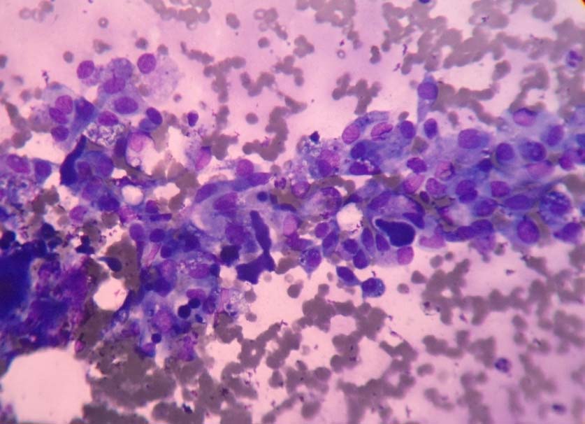
On re-evaluation certain characteristics like cellularity (mild, moderate or high), presence of follicular epithelial cells, MNGCs, epithelioid granulomas, colloid and inflammatory cells were taken into account. The most consistent feature seen in all the cases were the presence of follicular epithelial cells arranged mainly as honeycomb fragments, with fair number of these clusters showing degenerative changes [Table/Fig-1].
Follicular epithelial cells with degenerative changes along with thick colloid (MGG/40X).

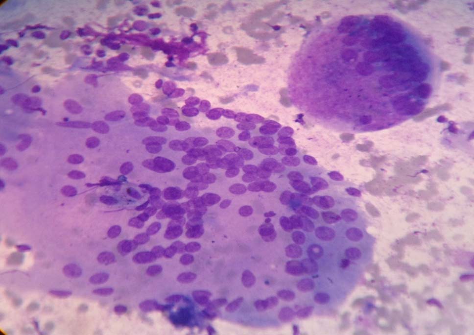
MNGCs were an invariable characteristic in all cases except one with the absence of MNGCs. Generally, these cells were scattered and occurred at high frequency with 2-3 MNGCs per low power field. The MNGCs were of variable size and shape, having multiple nuclei and abundant cytoplasm [Table/Fig-2]. Phagocytosed colloid and debris were also observed in the cytoplasm of some MNGCs.
A large multinucleated giant cell with abundant cytoplasm (MGG, 40X).

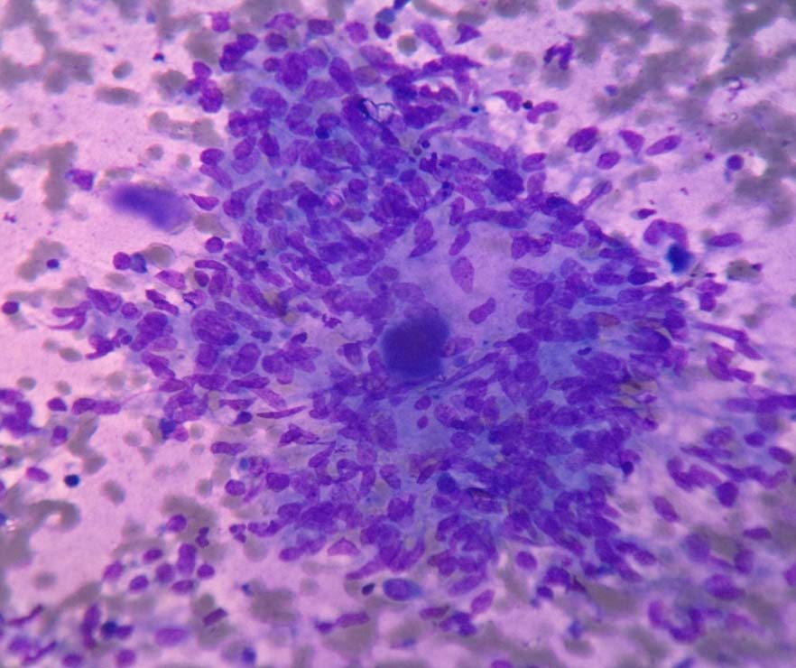
Scattered epithelioid cells and its clusters representing granulomas were observed in 15 of the total cases [Table/Fig-3]. They presented with variable morphology, ranging from loose to well formed granulomas.
Granuloma with ingested colloid (MGG, 40X).

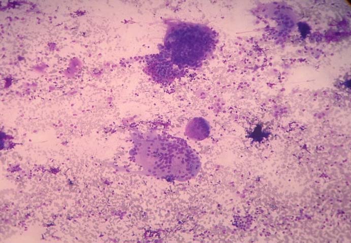
The other characteristic feature evaluated was the presence of colloid, which was observed occasionally only in 13 of the total cases. The colloid present was scant, darkly stained and thick, in variable, small and large forms scattered haphazardly [Table/Fig-1,4]. Occasional smear showed phagocytosis of the colloid by the MNGC’s and epithelioid histiocytes [Table/Fig-3].
Low power view showing multinucleated giant cells, follicular epithelial cells, colloid in a dirty background (MGG, 10X).

The features of dirty background of cellular debris, blood elements and pink amorphous acellular substance were observed in all 20 cases included in the study. Along with this a mixed type of inflammatory cells with predominance of lymphocytes and macrophages were also noted. It was observed that among 20 cases, nine cases showed presence of polymorpholeucocytes. The percentages of various cytological findings in the study are depicted in [Table/Fig-5].
Percentages of various cytological findings in the study.
| Cytological features | No of Cases (n=20) | % |
|---|
| 1 | Cellularity | | |
| Mild | 04 | 20% |
| Moderate | 16 | 80% |
| 2 | Follicular epithelial cells | 20 | 100% |
| 3 | Degenerated follicular epithelial cells | 13 | 65% |
| 4 | Multinucleated giant cells | 19 | 95% |
| 5 | Epithelioid granulomas | 15 | 75% |
| 6 | Dirty background and Inflammatory cells | 20 | 100% |
| 7 | Colloid | 13 | 65% |
The other thyroid diseases mimicking the clinical diagnosis were excluded by the absence of various cytological features like fire flares, oncocytic or hyperplastic changes. Papillary or microfollicular structures, plasmacytoid appearance, intranuclear inclusion or groove were not observed in any of the smears.
Discussion
DQT comprises nearly 3-6% of all thyroid lesions [1-3]. It is also called as granulomatous thyroiditis or subacute thyroiditis. The first case was diagnosed in 1825 and was labelled as “thyroiditis acuta simplex” till 1895. Later, in 1904 and 1936 this lesion were named as DQT after a Swiss surgeon De Quervain. DQT is a subacute, nonspecific inflammation of the thyroid gland whose aetiology is unknown. However similar to other viruses like mumps, hepatitis B and C, cytomegalovirus, enterovirus, and Type A and B coxsackie viruses the aetiology of DQT is also said to be virus related [2,4].
It usually appears several weeks after an upper respiratory infection and regresses within two to three months [2,5].
DQT is more common in females compared to males, the male to female ratio ranging from1:2 to1:6 [1,6]. However in our study the ratio was 1:1, with equal number of cases in both sex group.
Localised anterior neck pain, glandular tenderness and diffuse pain in ears are the usual clinical features of patients suffering from DQT. The other nonspecific features include fatigue, weight loss, low grade fever, elevated erythrocyte sedimentation rate, suppression in the thyroid stimulating hormone level and occasionally dysphagia [1-6]. In the present study, the commonest presentation was painful neck swelling.
In most of the cases, the level of C reactive protein is normal or slightly raised [2]. There is mild to moderate enlargement of the thyroid gland and it is tender on palpation [2,4,5]. The patients may be hyperthyroid, hypothyroid or euthyroid depending on the stage of the disease [1,2,4]. The recovery is usually spontaneous within weeks or months and may be supported with nonsteroid anti-inflammatory drugs or occasionally cortisone [1,7,8].
The diagnosis is usually made clinically when presenting with a typical history and fine needle aspiration is often not requested in such cases. It may clinically simulate neoplasm or a carcinomatous involvement of the thyroid gland and occasionally can be mistaken for a lymphnode near the thyroid. Thus, FNA is beneficial in the diagnosis of DQT and may provide very accurate results [9]. It also plays an important role to differentiate DQT from many other thyroid lesions mimicking it, specially malignancies and other forms of thyroiditis [1,3].
Fair number of MNGCs, epithelioid cell granulomas, inflammatory cells (lymphocytes, macrophages and neutrophils), degenerated follicular epithelial cells, and a dirty background composed of cellular debris, naked, degenerated nuclei and thick colloid are the key cytological characteristics for the diagnosis of DQT [1,8-11].
In the present study, the smears examined showed mild to moderate cellularity. All the 20 cases showed presence of follicular epithelial cells in sheets and clusters, out of which more than 60% of them showed degenerative changes. The follicular cells often show degenerative features and contain dark blue cytoplasmic granules representing lipofuscin or lysosomal debris. These granules are quite characteristic but are not specific for diagnosis and may be seen in involutional follicular cells of nodular goiter and occasionally in association with papillary and follicular neoplasms [12].
The giant cells are the hallmark of the disease [8]. In Our study 95% of the case showed presence of MNGCs as compared to 100% in study by Morde DA, Brachtel EF and Shabb NS et al., [5,13]. They are generally very large containing upto 200 nuclei, some containing ingested colloid. However, MNGCs often associated with lymphocytes are a common finding in papillary carcinoma, which should be considered in the differential diagnosis [3]. The other differentials to be excluded are granulomatous disease (sarcoidosis, tuberculosis and fungal infections) Hashimoto’s thyroiditis, Grave’s disease, palpation thyroiditis, and anaplastic carcinoma [6,13].
Another important feature is the presence of epithelioid granulomas. In our present study, 75% of the cases showed the presence of epithelioid granulomas against a dirty background comprising of mixed inflammatory cells with predominance of lymphocytes whereas in the study by Mordes DA showed epithelioid granulomas in 44.4% of cases [5]. In contrast the epithelioid granulomas in other granulomatous conditions are well formed, with fewer lymphocytes and absence of dirty background [2,12].
Colloid was present in 65% of the cases evaluated as compared to only 39% of the cases under study by Mordes DA [5]. In particular, presence of a non-foamy macrophage and monotonous cell reaction and epithelial cell degeneration are suggestive of the diagnosis of DQT.
All of the above features may not be observed in all the cases, and its presence depends on the stage of the disease at the time of aspiration. The characteristic cytological features may not be present in the resolving phase of the disease process. The absence of one or few of these findings does not exclude DQT, however diseases mimicking DQT in these cases have to be carefully excluded [3].
The presence of tumour cells with nuclear characteristics (powdery chromatin, nuclear groove and intranuclear inclusions), overlapping syncytial tissue fragments, real papillary fragments and psammoma bodies are characteristics in favour of Papillary Carcinoma of Thyroid gland (PTC). The classical MNGCs named as “bizarre giant cells” of PTC are smaller in number and size compared with DQT and granulomatous diseases. However, in some cases of papillary carcinoma, both type of MNGCs can be seen [6,13].
Limitation
The number of total subject considered in the study were 20, however future studies needs to be undertaken involving more number of cases along with hormonal and radiological correlation. The follow up of the patients were also not conducted as it was a retrospective study.
Conclusion
DQT is a self limiting inflammatory process of the thyroid gland. It presents clinically as a painful neck swelling, as observed in our study. They are often confused with other thyroid diseases and give rise to difficulties in diagnosis, hence FNAC proves to be an important tool in differentiating DQT from other thyroid lesions.
In conclusion, aspiration cytology of painful neck swelling showing follicular epithelial cells and MNGC’s against a dirty background is characteristic of DQT.