The Metabolic Syndrome (MS) is a multiplex risk factor for atherosclerotic cardiovascular disease. It consists of an atherogenic dyslipidemia, elevation of blood pressure and glucose, prothrombotic and proinflammatory states. The MS promote the development of cardiovascular disease at multiple levels [4,5].
There is approximately two fold risk of developing Cardiovascular Diseases (CVD) and fivefold risk of developing diabetes mellitus in individuals with metabolic syndrome. Depending on the number of components of MS present in individuals, the probability of developing diabetes and/or CVD within 20 years ranges between 30%–40% [6]. In South Asia 20%–25% of individuals have developed MS and many more may be prone to it [7,8]. Compared with other various distributions of adipose tissue in body, regional adipose tissue deposition is most critical in defining and initiating the metabolic syndrome. The visceral adiposity is associated with higher plasma glucose, high plasma insulin, hyperlipidemia and decreased HDL cholesterol concentrations which are components of metabolic syndrome [9]. Apart from biochemical and serological investigations, researchers are springing up with various imaging modalities to quantify and qualify the abdominal obesity.
The current precise and gold standard investigation for estimating the intra abdominal fat is Computed Tomography (CT) or Magnetic Resonance Imaging (MRI); however both are limited by their cost, availability and radiation hazards [3]. It has been advocated that ultrasonography can estimate the regional adiposity accurately and is easily available, safe and cost-effective [3].
This study was conducted to measure the regional adiposity of each patient suffering from metabolic syndrome by ultrasonography and correlating these measurements with biochemical factors, anthropometric measurements and body mass indices.
Materials and Methods
This was a cross-sectional study conducted in the department of radiodiagnosis and imaging KVG Medical College and Hospital Sullia, Karnataka, India, in subjects diagnosed with metabolic syndrome. Study was conducted from May 2013 to May 2015. Only patients with metabolic syndrome with age more than thirty years were included in the study whereas participants with no metabolic syndrome irrespective of body mass index and those who underwent recent abdominal surgery and liposuction were excluded from the study.
The study was approved by the Institutional Ethical Committee. All the subjects were informed about the study in their local language and written consent was taken from them.
After taking appropriate clinical history and recording blood pressure, height, weight and BMI of the patients, their Random Blood Sugar (RBS) values were obtained. Patients were diagnosed to have metabolic syndrome using the new International Diabetes Federation (IDF) criteria, which defines metabolic syndrome as a condition involving any three of the following parameters; regional obesity (measured in current study with BMI of more than 25 kg/mm2), raised triglycerides (>200 mg/dl), reduced HDL cholesterol (<35mg/dl), raised blood pressure (systolic> 140 mmHg or diastolic> 90 mm Hg) and raised fasting plasma glucose (>125 mg/dl) [10].
So in the current study, the three parameters were used for diagnosing metabolic syndrome viz., regional obesity (BMI > 25Kg/mm2), raised blood pressure (systolic> 140 mmHg or diastolic > 90 mm Hg) and raised RBS (>200 mg/dl). Only patients clinically diagnosed to have metabolic syndrome were included in the study. The body mass index (BMI) was calculated as weight in kilograms/height in m2. Blood pressure (BP) was measured by cuff method using a sphygmomanometer after keeping participant in resting position for at least 10 minutes.
During the entire study period, 105 subjects were clinically diagnosed to have metabolic syndrome using the above criteria and they were made to go through further thorough anthropometric measurements.
The patients clothing was removed from the waist line and was made to stand with feet shoulder width apart and back straight. Waist circumference was measured halfway between the lower rib and the iliac crest with the bottom edge of the measuring tape aligned with the top of the iliac crest and parallel to the floor. Measurement was done in full expiration. Similarly, hip circumference was measured at the level of the greater trochanter. The waist hip ratio was calculated [11].
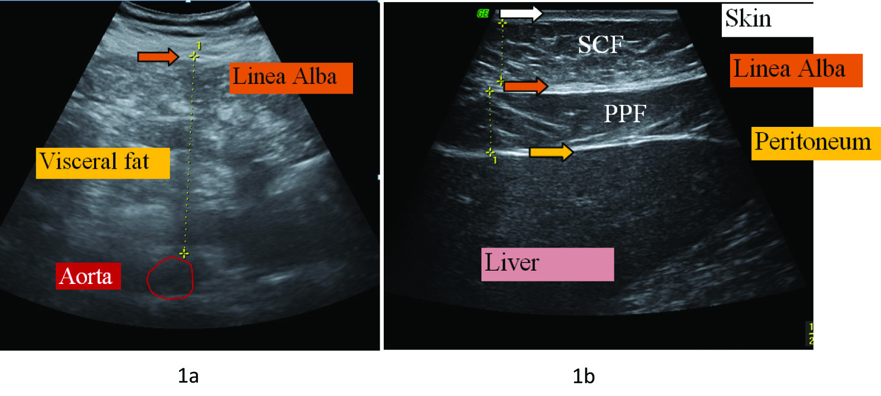
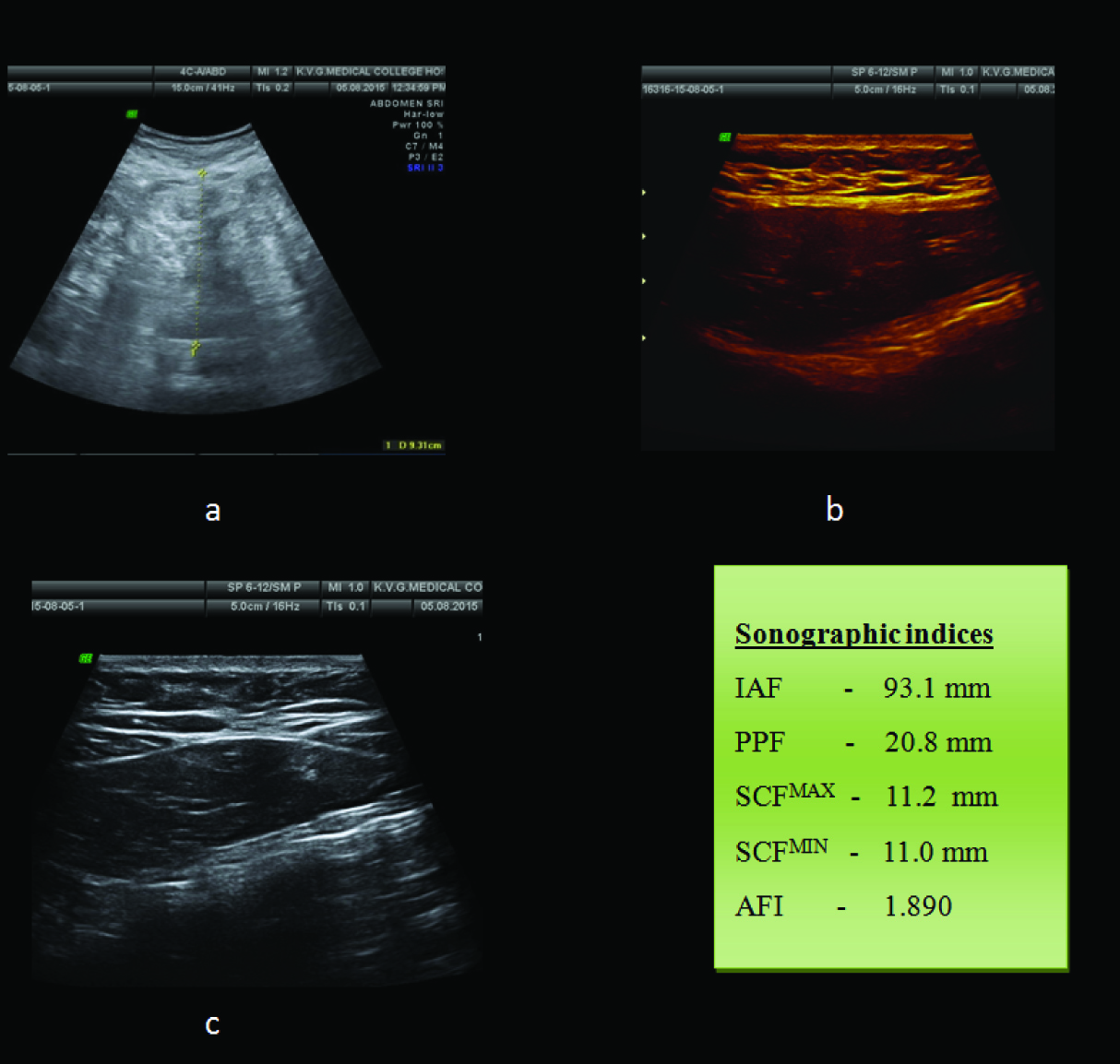
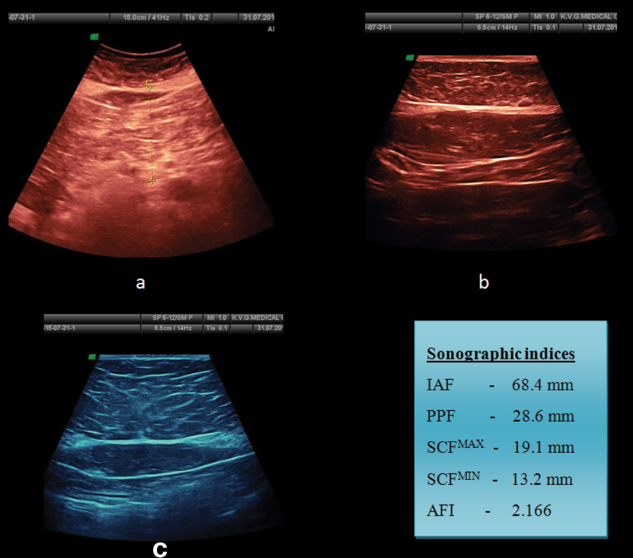
After recording anthropometric measurements trans-abdominal sonographies were performed with both curvilinear and linear transducers of GE Voluson 730 expert ultrasonographic machine. Patients were made to lie supine on ultrasound couch with relaxed abdomen and shoulders, heels, and buttocks in contact with the examination bed. Various sonographic indices were measured using either linear (7.5 MHz) or curvilinear probes (3.5 MHz) as follows [Table/Fig-1a,b, 2a-c, 3a-c].
a): Ultrasound abdomen with curvilinear (at supra umbilical region); b) Linear transducers (subxiphoid region) depicting technique of measuring intra-abdominal fat, minimum subcutaneous and maximum preperitoneal fat thicknesses.

a) Transverse view of epigastric region with curvilinear transducer demonstrating intraabdominal fat thickness; b) Longitudinal view of subxiphoid region with linear transducer depicting the measurements of minimum subcutaneous fat thickness and maximum preperitoneal fat thickness; c) Transverse view of epigastric region with linear transducer above the umbilicus demonstrating maximum subcutaneous fat thickness.

a) Transverse view of epigastric region with curvilinear transducer demonstrating intraabdominal fat thickness; b) Longitudinal view of subxiphoid region with linear transducer depicting the measurements of minimum subcutaneous fat thickness and maximum preperitoneal fat thickness; c) Transverse view of epigastric region with linear transducer above the umbilicus demonstrating maximum subcutaneous fat thickness.

Intra Abdominal Fat thickness (IAF): Measured with curvilinear probe in xiphoumbilical line mid way between xiphoid process and umbilicus. It is the distance between the anterior wall of the aorta and the posterior surface of the rectus abdominis muscle.
Preperitoneal Fat thickness (PPF): Measured in longitudinal plane with linear high frequency probe in the region of xiphoid process. It is the distance from the anterior surface of the liver to the posterior surface of the linea alba.
Minimum Subcutaneous fat thickness (SCFMIN): Measured in longitudinal plane with linear high frequency probe in the region of xiphoid process. It is the distance between the anterior surface of linea alba and the fat-skin barrier.
Maximum Subcutaneous fat thickness (SCFMAX): Measured in transverse plane with linear high frequency probe in the supra umbilical region. It is the distance between the anterior surface of linea alba and the fat-skin barrier.
Abdominal wall fat index (AFI): It is the ratio of maximum preperitoneal to minimum subcutaneous fat thickness [11].
Statistical Analysis
Statistical analysis were performed using SPSS (Statistical Package Social Science, version-10.0.5) software. Pearson correlation coefficients of anthropometry and sonographic indices with BMI were calculated which showed significant correlation at 0.05 level.
Results
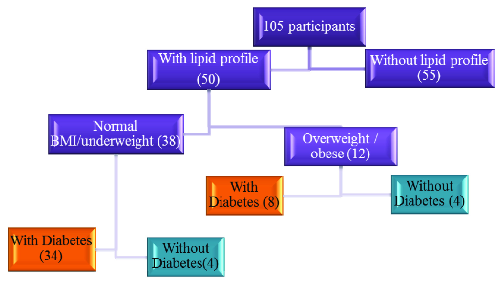
In the current study 105 patients were diagnosed to have metabolic syndrome by selecting three of following five criteria; obesity, raised triglycerides, reduced HDL cholesterol, raised blood pressure and raised fasting plasma glucose. Among them 48 were females and 57 were males. Subjects were distributed depending on their BMI [Table/Fig-4]. The distribution of the patients based on their BMI and presence of diabetes mellitus is portrayed in [Table/Fig-5].
Distribution of subjects based on BMI.
| Sl. No. | BMI categorization | Frequency |
|---|
| 1 | < 18 | Under weight | 5 |
| 2 | 18-24.9 | Normal | 33 |
| 3 | 25-29.9 | Overweight | 37 |
| 4 | >30 | Obese | 30 |
Distribution of participants.

The minimum age of the subject suffering from the metabolic syndrome was 31 years and maximum age of subject was 83 years. The mean age group in this study was 57.25 years with Standard Deviation (SD) of 11.23. Most of the metabolic syndrome cases in the present study were in the age group above sixth decade, followed by fourth and seventh decades.
According to one of the research conducted on Southern Indian population the cut off value of waist circumference for the diagnosis of central obesity in males is 85 cm and 80 cm in females [12]. With above reference values, 47 males and 44 females were categorized under central obesity. Same research work on Southern Indian population also provided the cut off value of waist hip ratio in males as 0.88 and in females as 0.81 [12]. With above reference values, 56 males and 47 females were categorized under central obesity.
Anthropometric measurements and sonographic indices were correlated with body mass index as standard. Pearson correlation coefficient was calculated at 2 tailed significance. The study showed positive correlation between BMI and waist/hip circumferences. Waist hip ratio showed poor correlation with BMI [Table/Fig-6]. Out of five sonographic indices AFI showed poor correlation with BMI. Intra-abdominal fat thickness, preperitoneal fat thickness, minimum and maximum subcutaneous fat thickness showed positive correlations with BMI [Table/Fig-7]. Minimum and maximum subcutaneous fat thicknesses showed much better correlation than others with r-value around 0.500. It was demonstrated that anthropometry is better for assessing the regional adiposity than sonographic indices. But indices like intra-abdominal fat, subcutaneous fat and preperitoneal fat thicknesses show higher association with BMI.
Pearson correlation coefficients of anthropometry with BMI.
| Parameters | BMI | WC | HC | WHR |
|---|
| Pearson correlation (r) | 1 | 0.624 (**) | 0.825 (**) | -0.115 |
| Significant (2 tailed) | | <0.001 | <0.001 | 0.254 |
| N | 105 | 105 | 105 | 105 |
Correlation is significant at 0.01 level.
BMI = Body Mass Index, WC = Waist Circumference, HC = Head Circumference, WHR = Waist Hip Ratio
Pearson correlation coefficients of sonographic indices with BMI.
| Parameters | BMI | IAF | PPF | SCFMIN | SCFMAX | AFI |
|---|
| Pearson correlation (r) | 1 | 0.324** | 0.211* | 0.585** | 0.513** | -0.207 |
| Significant (2 tailed) | | 0.001 | 0.035 | <0.001 | <0.001 | 0.039 |
| N | 105 | 105 | 105 | 105 | 105 | 105 |
Correlation is significant at 0.05 level.
Correlation is significant at 0.01 level.
BMI= Body Mass Index, IAF = Intra abdominal fat thickness, PPF = Preperitoneal fat thickness, SCFMIN = Minimum Subcutaneous fat thickness, SCFMAX = Maximum Subcutaneous fat thickness, AFI = Abdominal wall fat index
Considering the waist circumference as reference value for diagnosing the regional adiposity, its values were correlated with few significant sonographic indices like intra-abdominal fat thickness, minimum subcutaneous fat thickness and maximum subcutaneous fat thickness, which showed mild positive correlation with p-value of less than 0.05. A total of 105 participants were categorized into predominantly visceral adiposity if AFI is more than one and subcutaneous adiposity if AFI is less than one. A total of 40 participants had AFI less than one and referred as predominant subcutaneous adiposity where as 65 had AFI more than one and are referred as predominant visceral adiposity.
[Table/Fig-8] demonstrates the correlation coefficient values of anthropometric and sonographic indices for metabolic syndrome with random blood sugar levels. None of the anthropometric measurements showed positive correlation with random blood sugar, one of the metabolic risk factors. Whereas IAF, SCF and PPF revealed mild but positive correlation with random blood sugar levels.
Pearson correlation coefficient values of sonographic indices with random blood sugar levels.
| Parameters | RBS | IAF | PPF | SCFMIN | SCFMAX | AFI | Anthro pometry |
|---|
| Pearson correlation (r) | 1 | 0.0129 | 0.2015 | 0.0256 | 0.0444 | -0.1431 | -0.0272 |
| Significant (2 tailed) | | 0.900 | 0.047 | 0.802 | 0.664 | 0.160 | 0.0124 |
| N | 105 | 105 | 105 | 105 | 105 | 105 | 105 |
RBS = Random blood sugar, IAF = Intra abdominal fat thickness, PPF = Preperitoneal fat thickness, SCFMIN = Minimum Subcutaneous fat thickness, SCFMAX = Maximum Subcutaneous fat thickness, AFI = Abdominal wall fat index.
Discussion
This was a cross-sectional study performed on 105 consecutive individuals fitting into metabolic syndrome. Only 30 individuals in study were obese, 37 were overweight and remaining 38 were normal/under weight on BMI scale. However, they were included in the study as they had abnormal lipid profiles, diabetes mellitus and/or hypertension. Similar study was conducted by Kim SK et al., in Seoul, Korea where 32.65% were with normal BMI, 33.23% were overweight and 34.10% were obese [13]. They also included such individuals into the study as they were having abnormal lipid profiles, diabetes mellitus and/or hypertension. Hence, it is important to know that BMI is not the accurate judge of visceral adiposity, lean subjects categorized as normal or underweight may possess dreaded centrally located body fat which is responsible for catastrophe of metabolic syndrome [14].
Recently conducted study in Royapuram, Chennai, India, by Snehalatha C et al., gave cut off values for waist circumference as 85 cm and 80 cm for men and women respectively; correspondingly waist hip ratio of 0.88 and 0.81 respectively [12].
Following the same criteria we categorized our participants into centrally obese and non obese. By using above definition of waist circumference, 82.54% of males and 91.66% of females were classified as centrally obese. Waist hip ratio was still more significant and showed 98.24% of males and 97.9% of females as centrally obese in the current study.
Observing the [Table/Fig-9], it is clear that our study showed similar anthropometric results as that of studies conducted in Seoul [13], Taharan [15] and Netherland [16]. However, in the current study females had upper hand in waist circumference than males. In rest of the comparative studies males had more waist circumference than females. Waist hip ratios were almost similar in our study and in study conducted by Shabestari AA et al., [15].
Comparison of waist circumference and waist hip ratio of current study with various other studies.
| Studies | Waist circumference | Waist hip ratio |
|---|
| Male | Female | Male | Female |
|---|
| Current study | 92.64 ±11.72 | 96.95± 17.7 | 0.98 ± 0.14 | 0.92 ± 0.09 |
| Stolk RP et al., [16] | 93.9 ± 10.8 | 91.8± 9.5 | 0.91 ± 0.01 | 0.91 ± 0.01 |
| Kim SR et al., [13] | 88 ± 7.8 | 84.2 ±11.2 | 0.93 ± 0.04 | 0.93 ± 0.07 |
| Shabestari AA et al., [15] | 100 ±12.1 | 98 ±13.6 | 0.98 ± 0.05 | 0.90 ± .09 |
We correlated anthropometric measurements with BMI of participants. Waist circumference and hip circumference showed good correlation with BMI, with correlation coefficient values of 0.624 and 0.825 respectively. Waist hip ratio showed no correlation with BMI, with Pearson correlation coefficient of -0.115. Stolk RP et al., showed similar correlation with BMI and waist circumference with r-value of 0.824 [16]; whereas Shabestari AA et al., showed mild correlation with r-value of 0.285 [15]. Study conducted by Pouliot MC et al., concluded that waist circumference is much better anthropometric measurement than waist hip ratio and even better correlated with metabolic syndrome [17]. Authors also pronounced that waist circumference measured mid way between lower border of ribcage and iliac crest is more closely related with level of abdominal visceral adipose tissue than waist hip ratio in both sexes. Current study results almost marches on the same path of research conducted by Pouliot MC et al., and declares that waist circumference and hip circumference are better anthropometric indices for regional adiposity, where as waist hip ratio is poor indicator [17].
Next we compared and correlated various sonographic indices with BMI. Except for AFI rest four of sonographic indices showed positive correlation with BMI. SCFMAX and SCFMIN showed good correlation with BMI with correlation Coefficients of 0.513 and 0.585 respectively, followed by IAF with r-value of 0.324.
Maximum PPF depicted mild positive correlation with BMI with r-value of 0.211. Intra-abdominal fat thickness is considered as one of the most reliable sonographic index to measure visceral adiposity.
On sighting the [Table/Fig-10], it is clear that Kim et al., and Stolk et al., showed good correlation of IAF with BMI [13,16]. Current study gave comparable results as with researches conducted by Shabestari AA et al., Roopkala MS et al., and Shojaei MH et al., with r-value in the range of 0.32 to 0.46 [15,18,19].
Comparison of correlation coefficient value of IAF with BMI of current study with other studies.
| IAF with BMI | Current study | Stolk RP et al., [16] | Kim SK et al., [13] | Roopkala MS et al., [18] | Shabestari AA et al., [15] | Shojaei MH et al., [19] |
|---|
| Correlation coefficient value (r) | 0.324 | 0.640 | 0.610 | 0.395 | 0.436 | 0.462 |
On sighting the [Table/Fig-11] it was revealed that IAF compared and correlated with one of the metabolic risk factors (blood sugar levels), showed positive but mild correlation. In current study r-value was around 0.0129. Research conducted by other authors for the same showed r-values in the range of 0.1 to 0.3. So it can be concluded that IAF measured by sonography is better in estimating regional adiposity and shows mild positive correlation with metabolic risk factors.
Comparison of correlation coefficient value of blood sugar levels with IAF of current study with other studies.
| Blood sugar levels with IAF | Current study | Stolk RP et al., [16] | Kim SK et al., [13] | Seibert H et al., [22] | Shabestari AA et al., [15] |
|---|
| Correlation coefficient value (r) | 0.0129 | 0.190 | 0.310 | 0.100 | 0.239 |
Studies have shown good correlation between subcutaneous fat thickness and metabolic risk factors [20,21]. On scrutinizing the [Table/Fig-12] it is enlightening that subcutaneous fat thickness shows positive and good correlation with BMI in the current study. Studies done by Shabestari AA et al., and Roopkala MS et al., also show similar results [15,18]. Shojaei MH et al., showed very strong correlation between subcutaneous fat thickness and BMI with a value of 0.809 [19].
Comparison of correlation coefficient value of SCF with BMI of current study with other studies.
| SCF with BMI | Current study | Roopkala MS et al., [18] | Shabestari AA et al., [15] | Shojaei MH et al., [19] |
|---|
| Correlation coefficient value (r) | 0.585 | 0.677 | 0.510 | 0.809 |
On correlating blood sugar levels with minimum and maximum subcutaneous fat thicknesses in current study showed mild positive correlation with correlation coefficient values of 0.0256 and 0.044 respectively. Study conducted by Shabestari AA et al., showed likewise results with r-value of 0.009 [15]; whereas, Seibert H et al., showed poor correlation. Preperitoneal fat thickness is one of the better sonographic indices in diabetics and lean patients [22]. In lean patients subcutaneous fat thickness will be small resulting in high abnormal AFI, giving erroneous results [3]. Kim SK et al., and Shabestari AA et al., also depicted likewise results with r-value of 0.33 and 0.344 respectively [13,15].
While assessing metabolic syndrome risk factors in current study, preperitoneal fat thickness showed mild positive correlation with blood sugar values; r-value being 0.2015. Study conducted by Kim SK et al., showed similar results with r-value of 0.11. Among all sonographic indices preperitoneal fat thickness showed better correlation in assessing metabolic syndrome [13]. Tayama K et al., showed elevated preperitoneal fat thickness is associated with severity of diabetes mellitus, increased cardiovascular disease risk factors and poor prognosis in these participants [23].
In the evaluation of regional adiposity AFI showed poor correlation with BMI in the present study with correlation coefficient value of -0.115. Similar results were also obtained by Kim SK et al., and Shabestari AA et al., [13,15].
When scrutinized for the role of AFI for evaluating metabolic risk factors like blood sugar levels in the current study, it showed poor correlation with r-value of – 0.1431. In comparison studies performed by Kim SK and Shabestari AA revealed likewise results as ours with r-value of – 0.08 and 0.012 respectively [13,15]. In brief AFI is neither useful indicator for assessing regional adiposity nor metabolic syndrome risk factors.
Depending on AFI, participants are divided into two groups; prominent visceral fat depot with AFI > 1 and prominent subcutaneous fat depot with AFI < 1. Forty participants in the current study showed prominent subcutaneous fat depots and 65 participants with prominent visceral fat depots. No significant correlation was obtained when these two groups are compared with their RBS values. According to study conducted by Hashimoto M et al., individuals with prominent visceral fat depots are at increased risk of cardiovascular events than the individuals with subcutaneous fat depots [24].
Current study and study performed by Shabestari AA et al., reveals equivalent results when waist circumference is used as measurement tool for regional adiposity [15]. When waist circumference was evaluated for metabolic risk factors the current study lags showing no correlation, while study performed by Shabestari AA et al., shows mild positive correlation. In the current study none of the anthropometric measurements showed any sort of positive correlation with RBS values [15]. However, some sonographic indices like IAF, subcutaneous fat thicknesses and preperitoneal fat thickness showed mild but positive correlation with metabolic risk factors.
Limitation
Considering BMI as indicator for regional adiposity; even though all obese/overweight by BMI standards may not have increased regional adiposity and likewise all normal/lean individuals may not have decreased regional adiposity. Secondly, we compared only one of the metabolic risk factors (random blood sugar levels) with sonographic and anthropometric indices, as only less than half of participants were with lipid profiles. Finally, most of the participants were on regular treatment with antihypertensives, oral hypoglycemic drugs and antistatins.
Conclusion
Sonography can be considered as one of the reliable, easily available, repeatable, reproducible, cost effective and non ionizing imaging modality for assessing the regional adiposity but not as better as waist or hip circumferences. Raised sonographic indices like IAF, SCF and PPF are dependable markers of metabolic syndrome.
**Correlation is significant at 0.01 level.
BMI = Body Mass Index, WC = Waist Circumference, HC = Head Circumference, WHR = Waist Hip Ratio
*Correlation is significant at 0.05 level.
**Correlation is significant at 0.01 level.
BMI= Body Mass Index, IAF = Intra abdominal fat thickness, PPF = Preperitoneal fat thickness, SCFMIN = Minimum Subcutaneous fat thickness, SCFMAX = Maximum Subcutaneous fat thickness, AFI = Abdominal wall fat index
RBS = Random blood sugar, IAF = Intra abdominal fat thickness, PPF = Preperitoneal fat thickness, SCFMIN = Minimum Subcutaneous fat thickness, SCFMAX = Maximum Subcutaneous fat thickness, AFI = Abdominal wall fat index.