The gut wall displays a common structural plan which is modified region wise in accordance with the local functional differences. The general microstructure of gut is best appreciated by reference to its development. The alimentary canal originates as an endodermal tube enclosed in splanchnopleuric mesoderm. The endoderm forms the lining epithelium, secretory and ductal cells of glands of gastrointestinal tract. The splanchnopleuric mesoderm forms the connective tissue, muscle layers, blood vessels and lymphatics. The mature gut wall is comprised of four layers: mucosa, submucosa, muscularis externa and serosa or adventitia [1].
In the stomach, muscularis mucosa is composed of two thin layers of smooth muscles arranged as inner circular and outer longitudinal layer. The muscularis externa is three layered thick as outer longitudinal, middle circular and inner oblique layer and are oriented more randomly than layered [2].
The structural and functional knowledge of musculature of stomach along with its development is essential for the better understanding of pathogenesis of various associated gastric anomalies like hypertrophy of pyloric musculature and pathologies of nerve terminals and ganglia.
There have been earlier studies in this area on various animal foetuses, but there is paucity of literature regarding the histogenesis of stomach musculature in human foetus. Scant studies on human foetuses mostly include western population [3-7]. There are only two studies conducted on Indian foetuses [8,9] which show contrasting and conflicting views in the time of appearance of muscularis mucosa by the end of 22nd week [8] and 13 weeks [9] of gestation. While the western studies documented different time of development of muscularis mucosa at 18 weeks [5]. The exact time of development of the three concentric layers of muscularis externa is also debatable as it shows varying results in different studies [6-9].
Hence, the present study was conducted to assess the development of muscularis mucosa and muscularis externa in human foetal stomach.
Materials and Methods
The present observational study was conducted on 22 aborted human foetuses of gestational ages 10 to 26 weeks procured from the Department of Obstetrics and Gynaecology, Lok Nayak Hospital, Maulana Azad Medical College, New Delhi, India, from April 2007 to April 2010. The study was started after obtaining Institutional Ethical Clearance and written informed consent of the parent.
Procurement and Processing of Specimen
Three foetuses with gestational ages more than 24 weeks were obtained from spontaneous abortions and rest of the nineteen foetuses were below 24 weeks, obtained from cases coming for Medical Termination of Pregnancy (MTP).
The maternal status was assessed and an initial assessment of foetus was done to rule out any gross congenital abnormality. Hence foetuses with no gross congenital abnormalities were included in the study. The parameters as Crown-Rump length, Bi-parietal diameter and foot length were measured in all the procured foetuses. The gestational age of the foetuses were determined by evaluating the Crown-Rump length [Table/Fig-1].
Parameters used to determine the age of the foetuses collected.
| Gestational age (Weeks) | Crown Rump length (cm) | Bi parietal diameter (cm) | Foot length (cm) | Number collected |
|---|
| 10 | 6.5–7 | 2.2 | 0. 7–0. 8 | 2 |
| 12 | 8. 8–9.1 | 3 | 1.4–1.5 | 2 |
| 14 | 12.2–13.8 | 3.4 | 2.0–2.1 | 3 |
| 16 | 14.5–15.2 | 4.0–4.4 | 2. 7–2. 9 | 3 |
| 18 | 16.4–17 | 4.6–5.0 | 3.3–3. 4 | 3 |
| 20 | 19–20.1 | 5.0 | 4–4.1 | 3 |
| 22 | 21.2–21.5 | 5.2 | 4.2–4. 7 | 3 |
| 26 | 25–25.8 | 5.8 | 5–5.7 | 3 |
After procuring the foetus, a median incision was given on the anterior abdominal wall for immediate fixation of the gut. The foetus was then immersed for fixation in 10% formalin. After 24 hours, stomach was dissected and removed en-block from the lower end of oesophagus up to the pylorus and preserved in fresh fixative for two weeks. Specimens that showed any degree of autolysis were not considered for the study.
The dissected stomach was labeled and processed for paraffin embedding keeping the long axis of the stomach as the cutting surface. Seven micron thick serial sections were generated on a rotary microtome. Each longitudinal tissue sections had the gastro-oesophageal junction, cardia, fundus, body and pylorus.
Staining
Stomach sections of each gestational age were stained sequentially with Haematoxylin and Eosin to observe all the four layers of stomach. The structure, orientation and sequence of appearance of smooth muscle fibers was observed and studied in the developing stomach. Special stain, Masson’s trichrome was used to distinctly observe the muscle layers and differentiate it from connective tissue of lamina propria and submucosa.
The sections were then observed under a BX61 motorized microscope and the images were captured with Olympus DP71 camera. Processing of images was done with Image Pro plus MC 6 software and analysed.
Results
The present study evaluated the histogenesis of stomach muscularis externa and muscularis mucosa in the human foetuses ranging from gestational ages 10 to 26 weeks. This is a baseline study where we have been able to observe the microscopic structural details of the musculature of the entire foetal stomach, hence observing cardia, fundus, body and the pylorus.
As the age advanced, there was an increase in the size of the stomach. The layers of the stomach became more organized and were well distinct.
Fundus, Cardia and Body
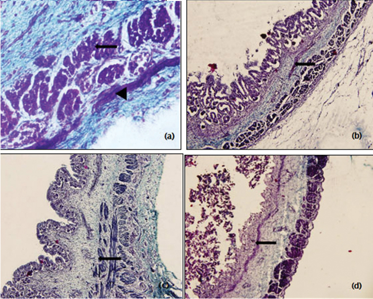
In the present study, the youngest foetus studied was of 10th week of gestation. During this period, all the layers of stomach were not well organized. The lamina propria was observed as a band of loose connective tissue just beneath the surface epithelium. The muscularis mucosa was not observed in any region of the stomach at this stage. Hence, the lamina propria was not distinct as a separate layer from the underlying connective tissue of submucosa. Outer to the submucosa, the muscularis externa was observed as a single layer of well defined circular muscle coat. While at some places, minimal discontinuous longitudinal smooth muscle fibers were seen external to the circular muscle coat [Table/Fig-2a].
Light micrograph of fundus (longitudinal section) stained with Masson’s Trichrome at different gestational ages; a) At 10 weeks (40X), showing the inner predominant circular muscle coat (arrow) and a discontinuous and thinner outer longitudinal muscle (arrowhead); b) At 14 weeks (10X), muscularis mucosa (arrow); c) At 16 weeks (10X), innermost oblique layer (arrow) of muscularis externa; d): At 22 weeks (10X), muscularis mucosa (arrow) seen as a continuous layer.

The smooth muscle fibers of muscularis mucosa were first evident at 14 weeks of gestation. These earliest strands of muscularis mucosa were observed as thin migratory radial strands of smooth muscle cells originating from the inner aspect of the muscularis externa towards the mucosal surface of the stomach and were stained eosinophillic with H&E and deep red with Masson’s Trichrome [Table/Fig-2b]. The lamina propria, at this stage was still not distinctive from the submucosa, as the muscularis mucosa although present was discontinuous. The muscularis externa at this gestation was two layers thick at some places, with a continuous layer of inner circular muscle coat, which was more predominant than the outer longitudinal muscle layer.
At 16 weeks, an additional discontinuous layer of oblique muscle fibers were observed to line the inner aspect of circular muscle coat of muscularis externa [Table/Fig-2c].
A continuous layer of well defined muscularis mucosa was observed at 22 weeks of gestation. The lamina propria and submucosa was thus well distinct as two separate layers at this stage [Table/Fig-2d].
At 26th week, the muscularis mucosa was thicker and continuous. Muscularis externa was also thickest and well defined into three distinct continuous strata as the outer longitudinal, middle circular and the inner oblique muscle layer. All the layers of stomach were most organized and resembled the adult picture.
Pylorus
In the pyloric part of developing foetal stomach, the muscularis mucosa was not observed at 10 weeks. Muscularis externa consisted of inner thicker predominant circular muscle coat with outer discontinuous longitudinal layer. Hence, four indistinct layers were observed as mucosa with merging lamina propria and submucosa, muscularis externa and serosa.
Muscularis mucosae appeared at the same time as in fundus and body at 14 weeks.
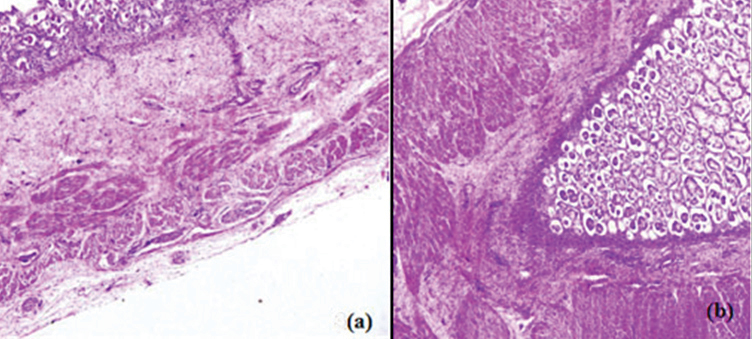
Muscularis externa was two layered thick with inner circular and outer longitudinal muscle coat from 10th week of gestation and became three layers thick with the advent of oblique muscle fibers being added inner to circular muscle fiber from 16 weeks. The muscularis mucosa became well developed and appeared as a continuous layer at 26th week. The Muscularis externa was also thickest and well defined at this stage as three continuous thick layers as inner oblique muscle, middle circular and the outer longitudinal layer. Muscularis externa was observed to be more developed and thicker than the rest of the region of stomach in the developing foetuses at all stages of gestation [Table/Fig-3a,b].
Light micrograph at 10X magnification stained with Haematoxylin and Eosin at 26 weeks of gestation: a) fundus; (b) pylorus.

Discussion
In the present study, histogenesis of the muscularis mucosa and muscularis externa was studied and the sequence and time of appearance of the various layers of stomach were established in the developing human foetus.
In our study, the muscularis mucosa was observed as thin and discontinuous strands of smooth muscle fibers, arising from the inner aspect of the muscularis externa towards the mucosal surface of the stomach at 14th week of gestation. However, earlier studies [5,8,9] show contrasting views regarding the time of appearance and staining pattern of muscularis mucosa. Arey LB observed muscularis mucosae in the foetuses at 160 mm CRL stage (18 weeks) [5]. The two most recent studies also differ in the time of appearance of muscularis mucosa by the end of 22nd week and 13 weeks of gestation [8,9]. At these stages, both the recent groups of researchers identified the smooth muscles of muscularis mucosa with H&E only. The muscularis mucosa was observed to be not differentiated and mature enough to take up Masson’s Trichrome at 22 weeks, but was distinctly stained by special stain Masson’s Trichrome at a late gestation by 28 weeks [8]. Whereas in our study we observed muscularis mucosa as early as in 14 weeks of gestation, which were stained well with both the H&E and Masson’s Trichrome.
Jirásek JE stated that in the human stomach, muscularis externa consists of circular layer from 10th gestational week, longitudinal coat being added from 11th week [6]. These findings were inconsistent with our study as we observed a continuous complete well developed circular muscle coat with thin longitudinal muscle layer by 10th week of gestation.
Maturation of gut smooth muscles were recognized in a rostro-caudal direction, from 8th week, with a large band of alpha-smooth muscle actin observed in oesophagus at week 8 and in the hindgut by week 11. The circular muscle was also documented to develop prior to longitudinal muscle [4]. This was in accordance with our study, where the circular muscle coat was more advanced in development than the longitudinal muscle coat.
Marciano T and Wershil BK documented that the development of muscular layer of stomach was complete by seven months [7]. We could not study foetuses above 26 weeks of gestation, hence cannot remark on the developmental status at later gestations.
Recent work on stomach musculature shows circular muscle layer from 15th gestational week, longitudinal layer being added by 28th week [8]. This is contradictory with our study where well developed circular muscle coat was observed with few scattered outer longitudinal muscle fibers as early as in 10th week. The same group of workers also observed the pyloric part with more extensively folded mucosa and deeper pits. The mucosal and muscularis externa layers were thicker in the pyloric part than in the body of the stomach [8]. This was in accordance with our study where the pyloric part was better developed and thicker than the body at all gestational stages of the foetuses.
The recent most study documented muscularis externa as well developed circular muscle layer with a thin layer of discontinuous longitudinal muscle fibres external to circular muscle coat at 10-12 weeks. The inner oblique layer was added from 16 weeks of gestation [9]. Similar pattern of development of muscularis externa was observed in the present study also with the circular muscle coat being continuous and predominant while discrete and discontinuous longitudinal muscle layer was observed outer to circular layer with no evidence of oblique muscle at 10 weeks. We also observed an additional discontinuous layer of oblique muscle fibers lining the inner aspect of circular muscle coat of muscularis externa at 16 weeks.
Thus in our study we were able to observe the exact time and sequence of development of muscle layers in muscularis mucosa and muscularis externa of each region of stomach at various gestational ages.
Limitation
The limitation of our study was the constricted range of gestational age of foetuses to near mid gestation from 10 to 26 weeks. Hence, we cannot comment upon how early the developmental changes must have appeared in the foetuses before 10 weeks. Rest all the foetuses were examined and studied in detail within the gestational age range of our study.
Conclusion
There is a sequential pattern of development and differentiation of muscularis mucosa and muscularis externa in stomach of human foetuses. The development of pylorus preceded the body and fundus portions of stomach in all foetuses at all gestational ages. This knowledge of embryological and developmental sequence in growth and development of musculature of stomach is significant in understanding the exact pathogenesis of various congenital anomalies like congenital pyloric hypertophy and associated anomalies of nerve and ganglia of stomach.