Pigmented Trichoblastoma of Nose: An Unusual Occurrence
D.C Sathyaki1, Mohammed Riyas2, Mereen Susan Roy3, R Jyothi Swarup4, Nibha Raghu5
1 Assistant Professor, Department of Ear, Nose and Throat, Sri Siddhartha Medical College, Agalakote, Karnataka, India.
2 Junior Resident, Department of Ear, Nose and Throat, Sri Siddhartha Medical College, Agalakote, Karnataka, India.
3 Assistant Professor, Department of Ear, Nose and Throat, Sri Siddhartha Medical College, Agalakote, Karnataka, India.
4 Professor, Department of Ear, Nose and Throat, Sri Siddhartha Medical College, Agalakote, Karnataka, India.
5 Junior Resident, Department of Ear, Nose and Throat, Sri Siddhartha Medical College, Agalakote, Karnataka, India.
NAME, ADDRESS, E-MAIL ID OF THE CORRESPONDING AUTHOR: Dr. D.C Sathyaki, Assistant Professor, Department of Ear, Nose and Throat, Sri Siddhartha Medical College, Agalakote-572107, Karnataka, India.
E-mail: sathyaki_dc@yahoo.co.in
Nevus sebaceus of Jadassohn is a congenital tumour affecting the scalp and face. It is usually presented as a pigmented patch or plaque. It is a complex cutaneous hamartoma which involves pilosebaceous follicle, epidermis and adnexal structures. Tumours that arise from nevus sebaceous are basal cell carcinoma, syringocystadenoma papilliferum, trichoblastoma and hidradenoma. The progression and frequency of the tumour increase with the age. Here we present a case of pigmented trichoblastoma over the external nose. It was a case of an elderly woman presenting with a painless swelling over the external nose which was soft, non-tender and with well defined margins.
Nevus sebaceous of Jadassohn, Trichoepithelioma, Tumour
Case Report
A 65-year-old elderly woman was presented with swelling over the external nose since one year, which was progressive, not associated with pain and no history of sudden increase in size.
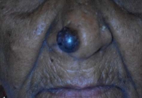
On examination, a pigmented plaque was found over the right side of the ala of the nose. It was 1x1 cm in dimension and was circular in shape. It was soft, non-tender with no local rise of temperature. Margins were well defined [Table/Fig-1].
The clinical presentation.
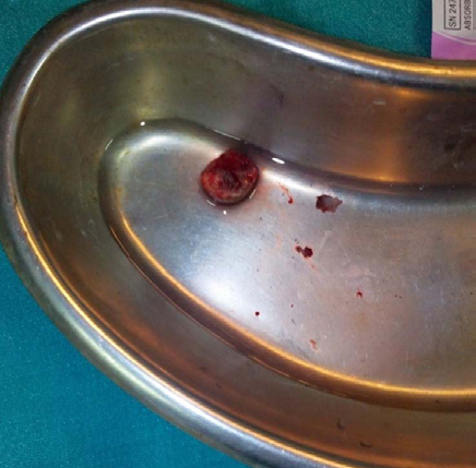
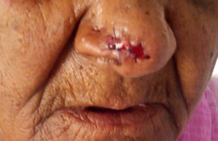
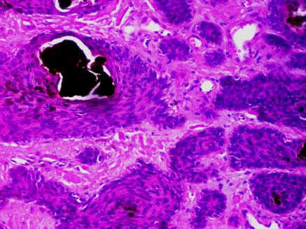
A wide excision with primary closure was performed [Table/Fig-2,3]. Histopathological examination showed cells arranged in nests and were separated by a fibrous stroma [Table/Fig-4]. Cells were round to oval, small (basaloid cells) and spindle shaped with elongated nuclei (mesenchymal cells) [Table/Fig-5]. Melanin pigment deposition was seen. Areas of necrosis with calcification were also seen [Table/Fig-6]. Stratified squamous epithelium with keratin was seen. Features were suggestive of pigmented trichoblastoma. Patient was followed up once in three months for two years and there was no recurrence.
The postoperative picture.
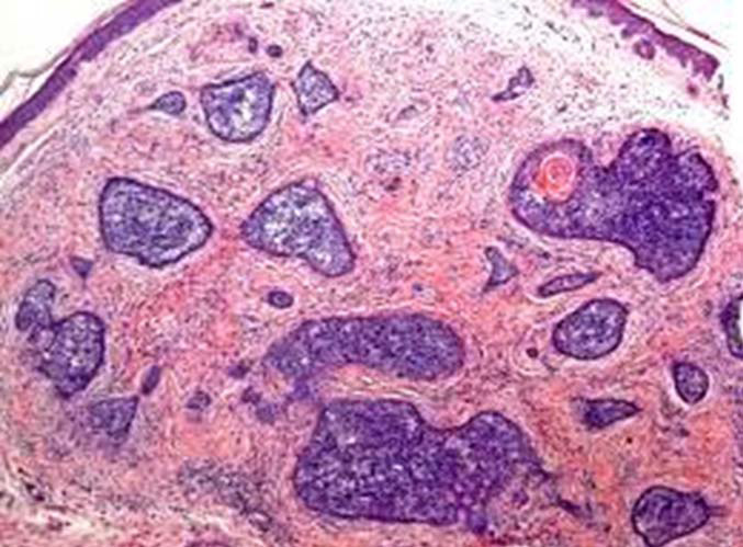
Histopathological section under 4X, showing a tumour in dermis which is separated from the epidermis.
Histopathological section under 10X, showing nests of basaloid cells with peripheral palisading separated by myxoid matrix.
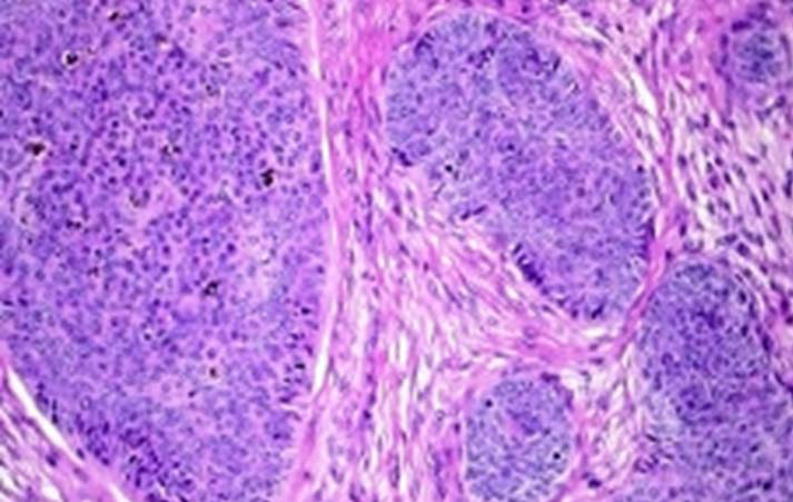
Histopathological section under 40X, showing nests of basaloid cells separated by myxoid matrix. Pigmentation is seen within the basaloid cells.
Discussion
Nevus sebaceous commonly appears as a yellowish patch or plaque at birth or in early childhood. Several benign and malignant tumours can develop from nevus sebaceous, with trichoblastoma and syringocystadenoma papilliferum, the most common benign forms. Trichoblastoma occurs as a primary or a secondary dermal neoplasm. It presents as a solitary, small, non ulcerated nodule over the scalp or face. Trichoblastoma is divided into five histological patterns. They are: large nodular (including pigmented), small nodular, cribriform, racemiform and retiform. Less common forms like adamantoid, columnar, rippled pattern, subcutaneous and superficial forms are variants [1]. Pigmented variants of trichoblastoma are very rare. Only few cases are reported in literature [1-3]. This is one such case of pigmented trichoblastoma. Trichoblastoma (trichoblastic fibroma) is located on trunk and scalp [4].
The histologic findings are basaloid proliferation in which tumour cells are arranged in cords, sheets, or discrete clusters surrounded by fibrous stroma. Pigmented trichoblastoma contains melanin deposits but no significant melanocytic hyperplasia [1]. Epidermal connection, atypia or mitosis is absent. Clefts are absent at stromal interface [5]. Nodular basal cell carcinoma and trichoepithelioma must be differentiated histologically. In this case, basaloid and mesenchymal cells were seen arranged in nests and was separated by a fibrous stroma. Myxoid stroma and stromal retraction or clefting around basaloid islands were absent which were characteristic of basal cell carcinoma [1]. Absence of keratotic cysts ruled out trichoepithelioma [6]. Pigmented trichoblastoma can be a part of a rare syndrome called phacomatosis pigmentokeratotica which is caused due to mutation of HRAS G123R gene [6].
Gozel S et al., reported a case of six tumours in the nevus sebaceus of Jadassohn in a single patient which included pigmented trichoblastoma. Other tumours were syringocystadenoma papilliferum, tubular apocrine adenoma, sebaceoma, tumour of follicular infundibulum and superficial epithelioma. The lesion was located in the scalp [1]. Cho HK et al., also reported a case of pigmented trichoblastoma in the scalp [2].
Here we present this case as the second case of pigmented trichoblastoma in the external nose. Kamat G et al., reported the first case of pigmented trichoblastoma over external nose [3].
Conclusion
Pigmented trichoblastoma is a benign neoplasm for which periodic follow up is necessary. If secondary neoplasms are developing then surgical excision is essential. Recurrence is uncommon.
[1]. Gozel S, Donmez M, Akdur NC, Yikilkan H, Development of six tumours in a sebaceus nevus of Jadassohn: Report of a caseKorean J Pathol 2013 47:569-74. [Google Scholar]
[2]. Cho HK, Song JS, Kang WH, Ro BI, Pigmented trichoblastoma arising from the nevus sebaceous: a rare case in KoreaAnn Dermatol 2009 21(4):406-08. [Google Scholar]
[3]. Kamat G, Yelikar B, Shettar S, Karigoudar MH, Pigmented trichoblastoma with sebaceous hyperplasiaIndian J dermatol Venereol Leprol 2009 75(5):506-08. [Google Scholar]
[4]. Krishnamurthy J, Divya KN, The cytology of giant solitary trichoepitheliomaJ Cytol 2010 27(3):99-101. [Google Scholar]
[5]. Fransisco GG, Said MC, Aldo GL, Gloria PV, Rodrigo VR, Rapidly growing pigmented tumour on a scalp nevus sebaceous of a pediatric patient: Observation or excisionDermatol Online J 2014 20(7):01-04. [Google Scholar]
[6]. Li JY, Berger MF, Marghoob A, Bhanot UK, Toyohara JP, Pulitzer MP, Combined melanocytic and sweat gland neoplasm: cell subsets harbor an identical HRAS mutation in phacomatosis pigmentokeratoticaJ Cutan Pathol 2014 41(8):663-71. [Google Scholar]