Fistulating Richter’s Hernia of Groin with Necrotizing Soft Tissue Infection: A Lethal Combination
Shibojit Talukder1, Ashish Gupta2, Basant Narayan Singh3, Lileswar Kaman4, P Abhinaya Reddy5
1 Senior Resident, Department of General Surgery, PGIMER, Chandigarh, India.
2 Senior Medical Officer, Department of Hospital Administration, PGIMER, Chandigarh, India.
3 Senior Resident, Department of General Surgery, PGIMER, Chandigarh, India.
4 Professor, Department of General Surgery, PGIMER, Chandigarh, India.
5 Senior Resident, Department of General Surgery, PGIMER, Chandigarh, India.
NAME, ADDRESS, E-MAIL ID OF THE CORRESPONDING AUTHOR: Dr. Lileswar Kaman, Professor, Department of General Surgery, PGIMER, Chandigarh-160012, India.
E-mail: drashish0403@yahoo.in
Strangulation of groin hernia can result in significant morbidity and mortality. Spontaneous external fistulation following strangulation is rare and typically occurs with Richter’s hernia. Spreading Necrotizing Soft Tissue Infection (NSTI) secondary to Enterocutaneous Fistula (ECF) is an ominous sign, further worsening its prognosis. Early diagnosis and prompt surgical treatment is crucial to improve outcome. Herewith the authors are presenting a case of neglected inguinal hernia. It was complicated with ECF formation and rapidly spreading NSTI of flank. He underwent resection and anastomosis of the gangrenous bowel, anatomical repair of the hernia along with soft tissue debridement of flank region. This patient however succumbed to sepsis with multi organ dysfunction. Significant delay in seeking medical care led to dismal outcome.
Enterocutaneous fistula, Metronidazole, Strangulated hernia
Case Report
A 60-year-old male presented to the emergency department with complaints of discharge of fecal matter from right groin for last three days. He had history of a reducible swelling in the same groin for past two years. It was asymptomatic so he never sought clinical attention earlier. However, for last one month the swelling was irreducible. For last seven days he was having progressively increasing dull aching pain in the region of the swelling along with redness over the swelling that gradually extended up to right loin. For last three days the swelling ruptured to produce two openings which started discharging feces.
There was history of constipation, obstipation, multiple episodes of bilious vomiting and progressive abdominal distension during last week. There was no history of fever, respiratory distress and decreased urine output. Patient was a heavy smoker for last for decades and used to take one quarter alcohol every alternate day. He was an unskilled laborer by profession.
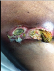
On examination patient was conscious and oriented. He had pulse rate of 110 beats per minute, blood pressure of 100/60 mmHg, respiratory rate of 24/minutes. He was afebrile and examination of respiratory and cardiovascular system was unremarkable. His abdomen was distended and nontender with sluggish bowel sounds. Examination of right inguinal region revealed a 10 x 5 cm ovoid swelling above and medial to pubic tubercle with over lying erythematous skin [Table/Fig-1]. Two ulcers were present on the swelling measuring 5 x 3 cm and 3 x 3 cm with undermined edges and sloughing base. There was feculent discharge from them. Swelling had no cough impulse. Left groin and genitalia were unremarkable. Note was made of a large area of erythema with induration and raise temperature extending from groin to left flank. There was blackish discoloration in the center of the erythema. Per rectal examination revealed fecoliths [Table/Fig-1].
Photograph showing fistulating right inguinal hernia with erythema and blackish discolouration of the flank
A clinical diagnosis of strangulated right inguinal hernia complicated by Enterocutaneous Fistula (ECF) and Necrotising Soft Tissue Infection (NSTI) was made. Patient was resuscitated with intravenous fluid, broad spectrum antibiotics (piperacillin-tazobactam and metronidazole) was started. Nasogastric tube was inserted and urinary bladder catheterization was done. His blood investigations revealed leukopenia (total leukocyte count 3700/mm3), raised urea and creatinine (urea 70 mg/dl, creatinine 3.6 mg/dl), metabolic acidosis. Chest radiograph was consistent with changes of chronic smoking.
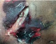
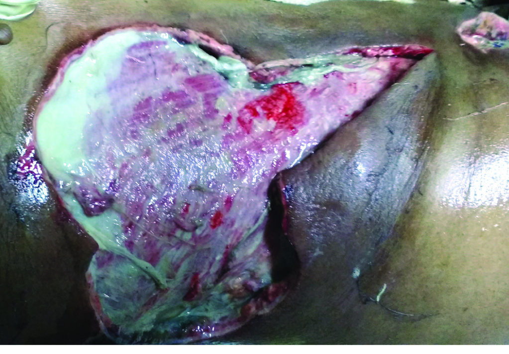
After adequate hydration and stabilization we look up for surgery under general anesthesia. The groin wound was explored first and it was found to have a loop of transverse colon along with part of the circumference of an ileal loop. The three fourth circumference of the entrapped ileal loop had gangrenous changes with perforation. Spillage of contents in inguinal canal was extensive. Suspecting intraperitoneal contamination a decision to explore the peritoneal cavity was made via midline laparotomy. We subsequently resected mid ileal segment and performed end to end anastomosis. There was no peritoneal contamination and transverse colon was healthy. The groin wound was debrided and anatomical facial closure was done with absorbable suture. Flank wound was incised over the black patch which revealed 20 ml of dish water pus and underlying soft tissue slough [Table/Fig-2]. An aggressive debridement over 15 x 10 cm area of the right flank was carried out [Table/Fig-3]. Patient was returned to surgical recovery on ventilator due to persistent metabolic acidosis.
Intraoperative image of necrotic patch with dish water pus.
Photograph of post debridement status of flank patch.
In postoperative period his surgical wound remained unhealthy with copious purulent discharge requiring further debridement. He was extubated on postoperative day 1. His renal function tests did not improve after surgery. On postoperative day 5 he developed progressive hypoxia and needed reintubation and positive pressure ventilation. There was bilateral course crepitation in chest and chest x-ray showed bilateral infiltrates. Subsequently he developed refractory hypoxia and hypotension and anuria and died on same day. The wound tissue culture showed growth of anaerobic organism sensitive to metronidazole.
Discussion
Hernias are one of the most common and oldest entities encountered in surgical practice [1]. Prevalence of ventral hernias is around 1.7% with inguinal hernia comprising approximately three quarter of them [2]. About 4% groin hernias present as surgical emergency [1]. Strangulation is the most catastrophic complication occurring in untreated cases. The incidence of strangulation varies from 0.29% to 2.9% [1]. Overall 30 day mortality from acute hernia surgery is approximately 0.02% and 0.48% respectively for below and above 60 years of age [3]. Mortality reaches up to 9% in presence of strangulation [1]. Incidence of strangulation and risk of morbidity and mortality are higher in old age.
Though Richter’s hernia was first described by Hildanus (1598), in 1897 Sir Frederick Treves credited August Gottlob Richter for providing first scientific report of the entity (1778) [4]. In Richter’s hernia partial circumference of a hollow viscus is entrapped in a relatively narrow hernial ring and undergoes rapid strangulation without compromising lumen. Though commonest to occur in femoral region (36-88%), it is possible to encounter them at other sites like inguinal region (12-26%), incision site (4-25%), drain site, laparoscopic port site, sacral foramina and Spigelian line [5-7]. They represent about a tenth to one third of all strangulated hernias. Review by Steinke W and Horbach JM showed that more than two third of theses hernias when strangulated develop rapid infarction [4,8].
Untreated strangulation with bowel obstruction is incompatible to life as rapidly ensuing metabolic jeopardy and septicaemia due to bowel gangrene leads to death [9]. Gillespie RW et al., while classifying Richter’s hernia found that the similar catastrophe may not occur in certain cases of Richter’s hernia where the necrosed bowel wall communicates with the atmosphere creating an ECF [10]. According to more recent classification depending on extent of bowel circumference getting strangulated a type I Richter’s hernia with less than one third bowel circumference will not produce any obstructive features while those with more than one third and two third circumferential involvement will result into incomplete and complete obstruction respectively [11]. Our patient had a type III hernia with three fourth of its circumference becoming gangrenous.
Fistulation of strangulated inguinal hernia into scrotum in pediatric and adult population has been recently reviewed by Ahi KS et al., [5]. They are predominantly reported from the developing counties with poor medical awareness and access to health services. Other sites like inguinal region [5,6], labia majora in females [12], femoral triangle [13,14] and umbilicus [15] has been cited sporadically. However coexistence of NSTI along with such fistulae is extremely rare as in present case. Our patient did not report immediately after fistula formation as his obstructive symptoms abated. This led to continuous and uncontrolled seepage of enteric contents into the surrounding soft tissue a known risk factor for NSTI. Similar case reports of NSTI complicating strangulated hernia sites like scrotum [16,17], groin [18], laparoscopic port site [19] umbilicus [20] and thigh [21] have been reported in literature. However involvement of the flank was unique to the present case.
All irreducible hernias with clinical suspicion of strangulation should be urgently explored [1]. A haemodynamically stable patient with preserved viability of strangulated viscus should undergo surgical reduction of strangulated hernia with anatomical or prosthetic repair depending on presence of contamination. Resection anastomosis is warranted in presence of compromised viability or frank infarction while prosthetic repair should be avoided [15]. Physiologically unstable patients may be best served with temporary ostomy creation as a part of damage control. NSTI when present merits extensive surgical debridement along with appropriate peri operative antibiotic cover [21]. Event with best treatment mortality rate of NSTI can be more than 80% [19].
Our patient died of multi organ failure due to sepsis. Acute setting surgery in strangulated hernia is associated with a sevenfold rise in mortality reaching almost 55% at 65 years [1]. According to literature a delay of more than 12 hours in strangulated hernia increases the chances of bowel resection [22]. In such event the mortality rate rises by 20 times [1]. Our patient was old, had presented late, underwent emergency surgery, needed bowel resection and also had necrotizing infection. All these factors synergized into a fatal outcome.
Conclusion
Strangulated Richter’s hernia is a rare surgical emergency. ECF with NSTI are result of extreme negligence in seeking timely medical attention. Prognosis in the presence of NSTI is dismal. Prompt diagnosis, early surgery with sepsis control measures can improve outcome.
[1]. Misiakos EP, Bagias G, Zavras N, Tzanetis P, Patapis P, Machairas A, Silvestro Canonico, Strangulated Inguinal Hernia, Inguinal Hernia 2014 InTechDOI:10.5772/57379 [Google Scholar]
[2]. Jenkins JT, O’Dwyer PJ, Inguinal herniasBMJ 2008 336(7638):269-72. [Google Scholar]
[3]. Bay-Nielsen M, Kehlet H, Strand L, Malmstrøm J, Andersen FH, Wara P, Danish hernia database collaboration. Quality assessment of 26 304 herniorrhaphies in denmark: a prospective nationwide studyThe Lancet 2001 358(9288):1124-28. [Google Scholar]
[4]. Steinke W, Zellweger R, Richter’s hernia and Sir Frederick Treves: an original clinical experience, review, and historical overviewAnnals of surgery 2000 232(5):710-18. [Google Scholar]
[5]. Ahi KS, Moudgil A, Aggarwal K, Sharma C, Singh K, A rare case of spontaneous inguinal faecal fistula as a complication of incarcerated Richter’s hernia with brief review of literatureBMC surgery 2015 15(1):67 [Google Scholar]
[6]. Shahbaz Habib F, Bushra S, Mohd Amanullah K, Afzal A, Syed Asmat A, Suprapubic faecal fistula due to richter’s inguinal hernia: a case report and review of literatureIran J Med Sci 2013 38(2):129-31. [Google Scholar]
[7]. Boughey JC, Nottingham JM, Walls AC, Richter’s hernia in the laparoscopic era: four case reports and review of the literatureSurg Laparosc Endosc Percutan Tech 2003 13:55-58. [Google Scholar]
[8]. Horbach JM, Invagination for Richter-type strangulated herniasTropical doctor 1986 16(4):163-68. [Google Scholar]
[9]. Friedman A, Strangulated Femoral Hernia complicated by Faecal FistulaMJ and Rec 1931 134:537-38. [Google Scholar]
[10]. Gillespie RW, Glas WN, Musselman M, Richter’s HerniaArch Surg 1956 73:590 [Google Scholar]
[11]. Kingsnorth A, The management of incisional herniaAnn R Coll Surg Eng 2006 88(3):252-60. [Google Scholar]
[12]. Elenwo SN, Igwe PO, Jamabo RS, Sonye US, Spontaneous entero-labial fistula complicating Richters hernia: Report of a caseInternational journal of surgery case reports 2016 20:27-29. [Google Scholar]
[13]. Kumar A, Pahwa HS, Pandey A, Kumar S, Spontaneous enterocutaneous fistula due to femoral herniaBMJ Case Reports 2012 2012:bcr2012006939 [Google Scholar]
[14]. Weledji EP, Puepi MA, Chichom AM, A rare spontaneous enterocutaneous fistulaJournal of surgical case reports 2014 2014(11)rju121 [Google Scholar]
[15]. Chen W, Liu L, Huang H, Jiang M, Zhang T, A case report of spontaneous umbilical enterocutaneous fistula resulting from an incarcerated Richter’s hernia, with a brief literature reviewBMC surgery 2017 17(1):15 [Google Scholar]
[16]. Onakpoya UU, Lawal OO, Onovo OD, Oribabor FO, Fournier’s gangrene compli-cating ruptured Richter’s inguinal herniaWest Afr J Med 2007 26:316-18. [Google Scholar]
[17]. Guzzo JL, Bochicchio GV, Henry S, Keller E, Scalea TM, Incarcerated inguinal hernia in the presence of Fournier’s gangrene: a novel approach to a complex problemAm Surg 2007 73:93-95. [Google Scholar]
[18]. Guirguis EM, Taylor GA, Chadwick CD, Femoral appendicitis: an unusual case. Canadian journal of surgeryJournal canadien de chirurgie 1989 32(5):380-81. [Google Scholar]
[19]. Losanoff JE, Richman BW, Jones JW, Trocar-site hernia complicated by NSTI—case report and review of the literatureHernia 2003 7(4):220-23. [Google Scholar]
[20]. Coyle P, Jaber S, Smith J, Grace PA, Damage control apronectomy for NSTI and strangulated umbilical herniaIrish journal of medical science 2010 179(4):607-08. [Google Scholar]
[21]. Georgiev-Hristov T, Álvarez-Gallego M, Juliá JB, Redondo MG, Verón A, Castell-Gómez JT, NSTI of the lower limb due to perforated inguinal herniaHernia 2011 15(5):571-73. [Google Scholar]
[22]. Tanaka N, Uchida N, Ogihara H, Sasamoto H, Kato H, Kuwano H, Clinical study of inguinal and femoral incarcerated herniasSurg Today 2010 40:1144-47. [Google Scholar]