Primary teeth are the best space maintainers and hence should be preserved and retained as long as possible [1].
Pulpectomy of primary teeth is indicated when the inflammation of the pulpal tissue involves the radicular pulp or when nonvital tooth is diagnosed. Pulpectomy helps in preserving a pulpally involved primary tooth by extirpating the diseased pulp associated with microorganism and debris from the canal and obturating with an antibacterial resorbable filling material [2].
The ultimate goal of pulpectomy is to achieve good hermetic seal which depends on various factors such as good biomechanical preparation, types of obturating material used and achievement of minimum voids. Obturation of the canal creates a fluid tight seal along the length of the root from the coronal opening to the apical system and eliminating all portals of entry between the periodontium and the root canal system [3,4].
Several different approaches to successfully fill the pulpectomized canals of primary teeth have been tried out. Cost effectiveness of carrier which is used to carry the material to the canal, ease of obturation, control and manipulation of material have been the key factors for successful outcome of clinically precise obturation. Different obturation techniques to fill the primary root canal include conventional manual incremental lateral condensation by amalgam pluggers, tuberculin syringe, disposable injection technique, navi tip, hand-held, rotary lentulospiral, jiffy tubes, endodontic pressure syringe, past inject, etc.
The lentulo spiral is the most commonly used instrument as the root canal paste carrier. The obturating paste can be filled by means of a manual lentulo spiral or mounted on micromotor handpiece. The process is easy and economical. Filling the root canal does not produce a densely compacted root canal filling and much reliance is placed on adherence of the paste to the walls of the canal [3].
Bhandari SK et al., used disposable syringe in their study. This is the simplest method of completely obturating canal space and filling the apical portion of the root canal and thereby, eliminating voids and incomplete filling along the root canal space [5].
The past inject paste carrier is a specifically designed device and works similarly to the lentulo spiral. The past inject provides good placement of the obturating material, while eliminating voids and providing a high density of the obturating materials [6,7].
So far, none of the obturation techniques available have been found ideal for obturation of root canals in primary teeth. Past inject is used for the placement of calcium hydroxide and root canal sealers in the permanent teeth, but there are not enough studies to evaluate its use as obturation technique in primary teeth. Thus, this study was undertaken to compare traditional techniques (i.e., lentulo spiral and disposable syringe) with past inject as obturation techniques in primary teeth. As one of the major cause of endodontic failure is incomplete obturation of the root canal system. Inadequate obturation of the tooth exposes it to periapical fluids, which provides material for growth of microorganisms or localization of bacteria in such dead spaces leading to subsequent sequelae of inflammation and ultimately endodontic failure. So the great emphasis needs to be placed on root canal filling materials as well as the technique of obturation. Various studies have been conducted to find out the ideal root canal filling material and best technique of obturation, but they all have been inconclusive.
Materials and Methods
The prospective study was approved by the Ethical Committee of the Government Dental College and Hospital, Jaipur, India, in accordance with 1975, Helinski declaration. The study population consisted of 41 healthy, cooperative children (four to nine years) with 60 teeth who had at least one primary tooth indicated for pulpectomy. The procedure and its possible discomfort and benefits were explained fully to the parents of the children involved and their written consent was obtained prior to the investigation.
Primary teeth requiring endodontic treatment, showing adequate bone support and root length (atleast two third root length remaining), with no radiographically detectable internal or pathological external resorption, were included in the study; patients having history of any systemic disease were excluded from the study. There were no drop outs in our study.
The overall 60 posterior teeth (120 canals) were randomly divided into three groups having 40 posterior canals for each group. The number of canals in the respective tooth was either three or four (during procedure) but due to two dimensional recordings in IOPAR and superimposition of canals, the visible canals (i.e., two canals mesial, distal) were assessed.
Before the start of the clinical study a full mouth dental examination and appropriate standardized periapical radiographs, were taken of mandibular posterior teeth with possible indication for pulpectomy. To obtain accurate radiographs the Rinn XCP instrument (Dentsply Rinn, Elgin) and radiographic parallelism and standard exposure technique were used to permit good visualisation of the tooth structure as well as reproducibility.
The primary tooth single sitting pulpectomy, was carried out by the same operator in all cases after administration of local anaesthesia and placement of a rubber dam. The procedure involved cavity preparation, removal of all carious tooth structure, preparation of a straight line access, extirpation of pulpal debris from the root canal using files, and copious irrigation with normal saline. Fine reamers were gently inserted into the canal and diagnostic radiographs were taken to establish the length of the root canals. The working length was maintained 1 mm short of the apex while preparing the canals with Hedstrom files (30-35 sizes) using a pullback motion. Care was taken to perform a selective filing, i.e., while filing the root canal, more pressure was maintained along the outer wall of the canal as the walls towards the inter-radicular areas are generally thin due to physiological resorption with associated risk of perforation. The root canals were thoroughly irrigated with sodium hypochlorite; saline solution was used as the last irrigating medium. The root canals were then dried using absorbent paper points inserting them 1 mm short of the radiographic apex. A homogenous mixture of zinc oxide and iodoform paste (Endoflas) was used according to the manufacturer’s instruction for filling the root canal using one of the randomly assigned assessed techniques.
Teeth in Group I were obturated with a disposable syringe (Dispovan), Group II using lentulo spiral (Mani) and teeth in Group III were obturated using past inject (Micromega).
The disposable syringe (Dispovan) (Group I) with a 25/26 gauge needle, 1.5-inch needle was taken, and the sharp edge of the needle was blunted using a sterile diamond point. The needle was placed 2 mm short of the radiographic apex and gradually material was expressed into the root canal. The apical end of the canal was filled first and by gradual withdrawal of the needle, the remaining canal space was subsequently filled.
The lentulo spiral (Group II) selected was two sizes smaller than the last file used and it was inserted 1 mm short of the root canal apex of primary teeth, thus reducing the risk of fracture. Lentulo spiral was hand held as there is no statistically significant difference between the manual and motor driven technique of obturation, according to the quality of the root canal filling or success rate [3]. Cotton pliers holding a wet cotton pellet were used to lightly press the material inside the canal, creating space for a temporary restoration. A similar technique was used for the past inject group (Group III).
Postoperative Evaluation
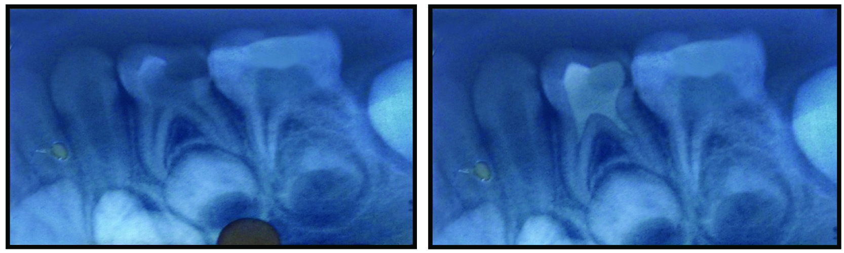
Postoperative IOPA radiographs of each tooth were obtained for each tooth using the same radiographic settings as for pre-operative [Table/Fig-1,2,3 and 4]. Radiograph of each tooth was examined to assess the quality of obturation.
a) Preoperative IOPA radiograph; b) Postoperative IOPA radiograph with half of root length obturated. (Images left to right)
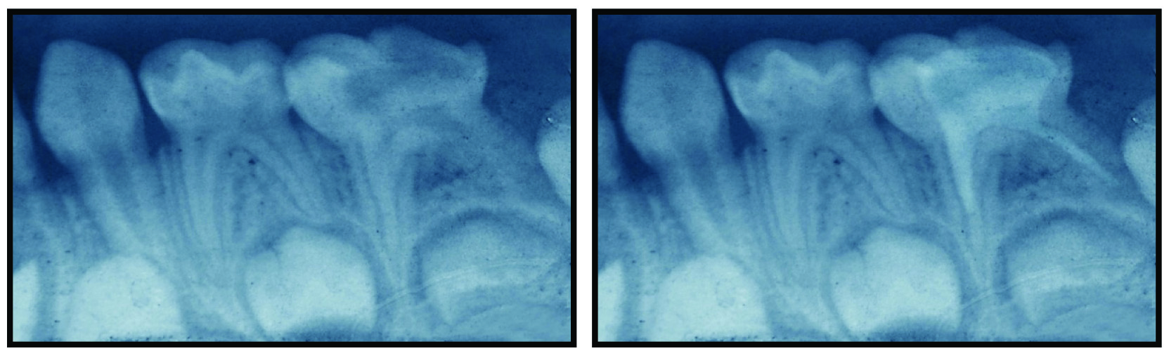
a) Preoperative IOPA radiograph; b) Postoperative IOPA radiograph with more than half of root length obturated. (Images left to right)
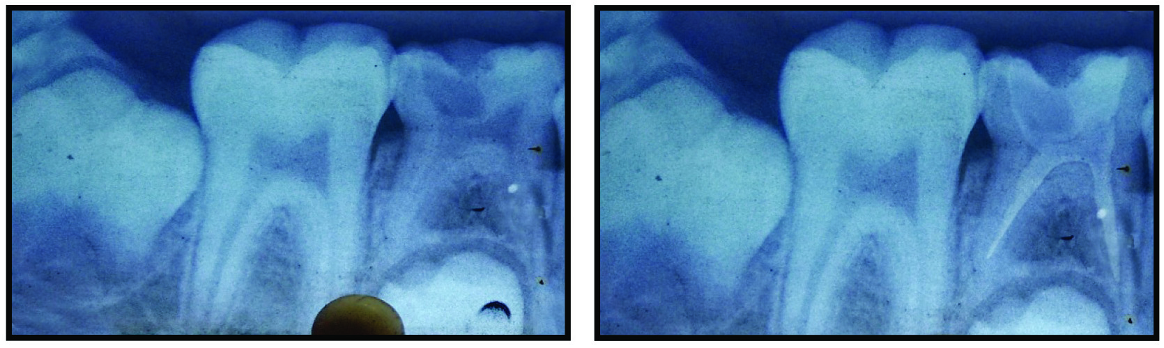
a) Preoperative IOPA radiograph; b) Postoperative IOPA radiograph with optimally obturated canal. (Images left to right)
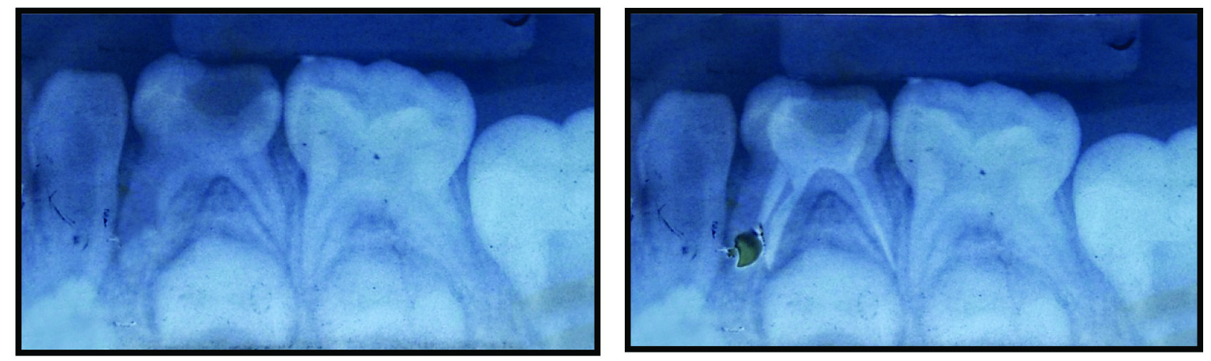
a) Preoperative IOPA radiograph; b) Postoperative IOPA radiograph with over obturated canal. (Images left to right)
For the molars, the mesial canals were considered as a single canal because of superimposition of the two canals seen on the intraoral periapical radiograph. Two evaluators, blinded to the filling technique and material, assessed presence of voids and obturation. Quality of obturation was scored as per scoring criteria given by, Memarpur M et al., [Table/Fig-1,2,3 and 4] [8].
Postoperatively radiograph of each tooth was evaluated by naked eye for the presence or absence of voids.
Statistical Analysis
The data was coded and entered into Microsoft Excel spread sheet. Analysis was done using SPSS version 20.0 (IBM SPSS Statistics Inc., Chicago, Illinois, USA) windows software program. The variables were assessed for normality using the Kolmogorov Smirnov test. Descriptive statistics included computation of means and standard deviations. Chi-square test used for qualitative data whenever two or more than two groups were used to compare. Level of significance was set at p=0.05. (p=<0.05 – significant, p=<0.01 – highly significant, p=<0.001 – very highly significant).
Results
A total of 13 females and 28 males with age ranging from four to nine years were enrolled in the study. All teeth were indicated for single sitting pulpectomy with no particular gender predilection. Postoperatively there was no sign of infection or any untoward reaction in any of the 60 teeth. The data were analysed to assess the success rate of the three methods used for obturation using Chi-square test.
Quality of Canal Obturation
Assessment of the root canals with different levels of obturation showed that Group II exhibited the highest number of underfilled canals (25%), while the highest percentage of optimally filled canals was observed in Group III (18.3%%), and the highest percentage of overfilled canals was observed in Group I (10%). Significant differences (p< 0.05) were observed when frequency for underfilled, optimally filled and overfilled canals was compared, respectively [Table/Fig-1,2,3,4 and 5].
Comparisons for different levels of obturation with various obturating techniques.
| Parameters | Scores | Total | p-value |
|---|
| Less or equal to half the root length (Score-1) | More than half the root length (Score-2) | Optimal filing (Score-3) | Filling extruding from the apex (Score-4) |
|---|
| Disposable syringe | N | 4 | 5 | 5 | 6 | 20 | 0.01 (S) |
| % | 6.7% | 8.3% | 8.3% | 10.0% | 33.3% |
| Lentulo spiral | N | 6 | 9 | 3 | 2 | 20 |
| % | 10.0% | 15.0% | 5.0% | 3.3% | 33.3% |
| Past inject | N | 0 | 7 | 11 | 2 | 20 |
| % | 0.0% | 11.7% | 18.3% | 3.3% | 33.3% |
| Total | N | 10 | 21 | 19 | 10 | 60 | |
| % | 16.7% | 35.0% | 31.7% | 16.7% | 100.0% | |
S=Significant Test applied: Chi-square test.
Voids
The highest percentage of voids were seen in canals filled with lentulo spiral (20%) minimum number of voids was observed in canals filled with past inject technique (6.7%) and pressure syringe (8.3%) [Table/Fig-6].
Percentage of voids in canals obturated using different methods.
| Parameters | Voids | Total | p-value |
|---|
| 0 | 1 |
|---|
| Disposable syringe | N | 15 | 5 | 20 | 0.01(S) |
| % | 25.0% | 8.3% | 33.3% |
| Lentulo spiral | N | 8 | 12 | 20 |
| % | 13.3% | 20.0% | 33.3% |
| Past inject | N | 16 | 4 | 20 |
| % | 26.7% | 6.7% | 33.3% |
| Total | N | 39 | 21 | 60 | |
| % | 65.0% | 35.0% | 100.0% | |
S=Significant Test applied: Chi-square test.
Discussion
Maintenance of an intact deciduous dentition until the eruption of the permanent successors is very important so as to maintain the arch form. Resolution of the pulpal infection with the help of endodontic treatment can preserve arch space as well as restore function. In addition to preserving arch form, utilization of pulp therapy to maintain the integrity of the primary dentition may also prevent aberrant tongue habits, prevent speech problems, maintain normal masticatory function and preserve aesthetics.
In literature there are plenty of studies to evaluate the success rate of different root canal filling materials used for primary teeth in comparison to obturating technique studies. So the present study was carried out to compare the clinical efficacy of three root canal obturating techniques in primary teeth.
The root canal therapy technique in this study was carried out on all teeth by a single operator. A study design with a single operator offers the advantage of consistent and reproducible technique. However, a potential limitation of this study design would be that the outcome is due in part to a superior operator rather than a superior technique. The technique itself is not standardized but is operator sensitive.
Pulp management of infected primary teeth involves not only thorough debridement of the root canal system but also obturation by using a material which is biocompatible and would resorb at the same rate as the roots of the involved tooth, without endangering the succedaneous permanent tooth and its eruption. Till date, a number of investigators have tested different materials but none of these have been shown to possess the requisite properties of an ideal root canal filling material and techniques for primary teeth, especially with regard to the major desirable property of having a rate of resorption matching that of the physiologic root resorption of the primary teeth.
In the present study, endoflas was used as obturating material. The material is hydrophilic and can be used in mildly humid canals. It firmly adheres to the surface of the root canals to provide a good seal. Due to its broad spectrum antibacterial activity it has the ability to disinfect dentinal tubules and difficult to reach accessory canals that cannot be disinfected or cleansed mechanically [9]. Endoflas resorbs at the same pace as the physiological resorption of root. This factor results in the resorption of the material limited to the excess extruded extraradicularly without showing any signs of resorption intraradicularly. Endoflas has high clinical as well as radiographic success of over zinc oxide eugenol [10].
In a retrospective study by Fuks AB et al., endoflas was used as a filling material. The resorption of the material was limited to the excess extruded extra-radicularly and it does not get depleted intraradicularly. The over pushing of the root canal filling material in primary teeth is unavoidable in some cases because of the thin dentinal walls of the root canals towards the inter-radicular areas, which may give way during filing of root canals [11].
In the present study, all techniques used to fill the canals led to voids in the filling material, a finding consistent with earlier reports [12-14]. The voids may create leakage in the paste, and thus may lead to microorganism regrowth, reinfection and an increased risk of post-treatment disease, especially if there are several large voids [13-17]. Factors that influence the location and size of the voids include the type, viscosity, and consistency of the paste, the method used to apply the paste, and operator skill and experiences [13,18].
The highest number of voids was seen in canals filled with the lentulo spiral (20%), and disposable syringe group (8.3%).
Dandashi MB et al., also found in their study that the disposable syringe system produced fewer voids as compared to the lentulo spiral [15]. Similar results were obtained by Oztan MD et al., who compared two different carriers for intracanal placement of calcium hydroxide: lentulo spiral and past inject. They observed that most of the canals filled with lentulo spiral showed a significantly higher number of voids compared to past inject [7].
The majority of the canals obturated with past inject showed optimal filling. Success of the technique can also be attributed to the fact that past inject is a specially designed paste carrier with flattened blades, which improves material placement into root canal, causing a lower occurrence of underfilled and overfilled canals as well as voids [6,7].
According to Grover R et al., past inject exhibited the highest number of optimally filled canals, and the highest number of overfilled canals were observed with pressure syringe [19]. Minimum number of voids were present in canals filled with the past inject technique and pressure syringe. These results suggest that past inject was the most effective technique for obturation of primary teeth.
The highest number of overfilled canals was observed with the disposable syringe. This might be due to the excessive pressure placed while pressing the material into the canal.
Available injection techniques illustrated in the literature have a universal defect, which is the overfilling of the obturating material that leads to inflammation and related complications [20]. The syringes have been proven to be better than the lentulospiral in controlling the voids and are superior in controlling overfills when used judiciously [12,13].
In present study, disposable syringe was used for delivering paste. As the technique achieves all the desired requirements of good root canal obturation, that is homogenous fill up to the desired depth as the obturation starts from apex upward, leaving no room for entrapment of air and formation of voids, the problem of apical overfills [5].
Nagar P et al., in their study used insulin syringes but still they obtained slightly more number of overfillings. Thus, they stated that optimum operator skills and proper material mix can provide optimal filling with less number of voids [21].
According to Parikh A et al., the lentulo spiral technique produced fewer extrusions [22].
In present study, sample size was not adequate to come to a final conclusion. The evaluation criteria in present study was IOPA radiographs which is a 2-D view so voids and filling are evaluated in 2-D view only. Further clinical trials with larger sample size and evaluation in 3-D (CBCT) are needed to validate the results of present study.
Conclusion
From the analysis of results and within the limits of this study the past inject technique proved to be the most successful, yielding a higher number of optimally filled canals and minimal voids, combined with easier placement of the material into the canals. Thus, this method appears to be of prospective interest for use in paediatric dentistry. The other tested methods disposable syringe, lentulospiral produced less acceptable results. In the literature, there are not sufficient investigations that support the use of past inject as an obturation technique for primary teeth. However, further clinical studies are needed to validate the results of the present study.