A Rare Case of Bi-rooted Deciduous Canine
Shrestha Shukla1, Aarti Garg2, Ravinder Garoo3
1 Senior Resident, Department of Dentistry, Clove Dental, New Delhi, India.
2 Assistant Professor, Department of Pedodontics and Preventive Dentistry, Jaipur Dental College, Jaipur, Rajasthan, India.
3 Senior Consultant, Clove Dental, New Delhi, India.
NAME, ADDRESS, E-MAIL ID OF THE CORRESPONDING AUTHOR: Dr. Shrestha Shukla, QU-45/B Pitampura, New Delhi, India.
E-mail: drshreshthashukla@gmail.com
Anomaly, Multirooted, Primary canine
A 10-years-old child was reported to our clinic by his parents with chief complaint of delayed eruption of permanent teeth in upper and lower jaws. There was no history of trauma or any hospitalization. On intra-oral examination, maxillary lateral incisors were missing.
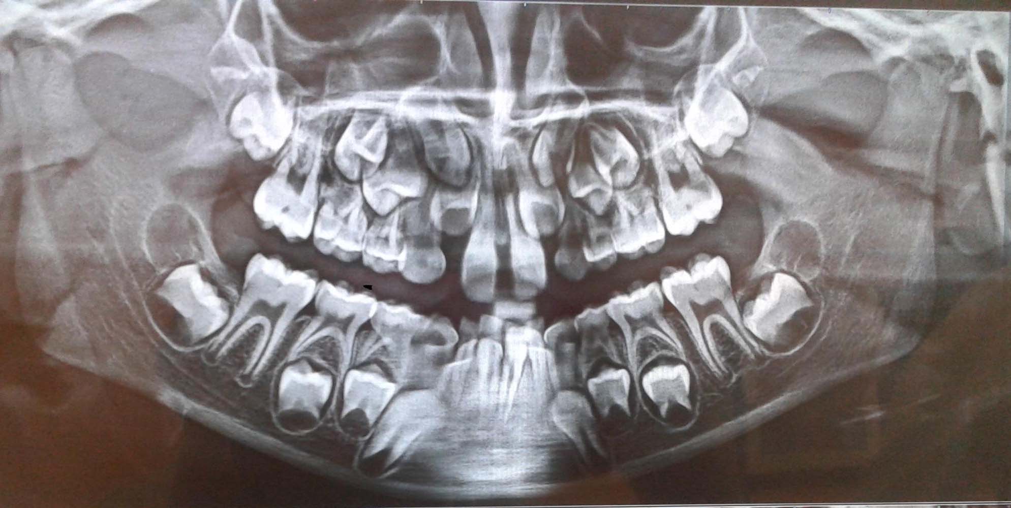
On panoramic radiographic examination [Table/Fig-1], the permanent maxillary lateral incisors were trapped between deciduous canines and permanent central incisors bilaterally. A decision was made to extract deciduous canines to allow removal of obstructions and obtain an easy path of eruption for lateral incisors after Bolton’s space analysis of maxillary arch. Left maxillary deciduous canine was extracted atraumatically under local anaesthesia (lignocaine with adrenaline 1:80,000). Patient was recalled after few days and the extraction of right maxillary deciduous canine, under local anaesthesia, was done.
Orthopantomogram showing bilateral deciduous canine.
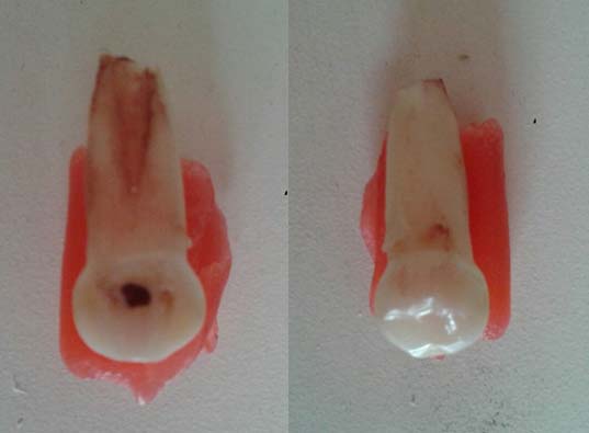
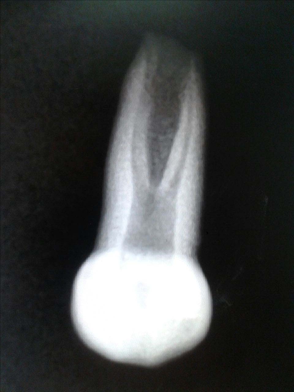
On examination of the extracted teeth, it was found that the extracted right maxillary deciduous canines had double root palatally and a single root was evident labially [Table/Fig-2]. On radiographic analysis of extracted teeth [Table/Fig-3] a wide pulp chamber and two separate roots were seen.
Extracted right maxillary deciduous canine- tooth was single rooted labially and palatally, it showed two roots.
Radiograph of the extracted deciduous canine showing wide pulp chamber and two separate roots.
In the permanent dentition, supernumerary roots are not uncommon, with normally single-rooted permanent premolars and canines being particularly affected [1].
The primary canines reported to date, demonstrate root furcations in the coronal third. Their bi-root formation, beginning between 9 and 10 months postnatally, may result from an enhanced expressivity of the gene initiating differential growth of Hertwig’s epithelial root sheath in multirooted teeth [2]. Although trauma or other disturbances in morphodifferentiation may affect root form and size in later periods, early trauma would not explain the appearance of bilateral birooted primary canines [2].
Although birooted primary canines cannot be detected by regular intra-oral examination, they may easily be observed by assessment of routine dental radiographs. It is important to diagnose such cases as this unusual root anatomy can lead to complications during endodontic procedures as well as during extraction of such teeth. Moreover, these anomalies of the deciduous teeth can impose problems in the eruption of succedaneuos teeth [3].
The mechanism for normal development of multiple roots is well known. From the cervical loop of the dental organ, the inner and outer enamel epithelia proliferate as a double layer of cells known as Hertwig’s epithelial root sheath. At the future cemento-enamel junction, the outer and inner enamel epithelia bend creating the epithelial diaphragm. The rim of this sheath encircles the primary apical foramen. An unknown factor triggers continued morphodifferentiation in multirooted teeth. By differential growth, tongue like extensions of the horizontal diaphragm develop, grow toward each other, and fuse. From each new secondary apical foramen, a root develops [4].
Birooted primary canines cannot be detected by routine intra-oral examination and sometimes can be missed by the clinicians. Although these primary canines may resorb and exfoliate without interference with eruption of the adjacent permanent teeth, but if noted, the anomaly should be described to the parents.
During exodontic procedures, the clinician should make sure that the crown of the permanent tooth is not trapped in the inter-radicular area of the primary tooth as this may cause accidental removal of the developing permanent tooth [5]. Here, it is suggested that, before extraction of a primary canine, this anomaly should be kept in mind and radiographs should be taken routinely before extraction.
[1]. Shafer WG, Hine MK, Levy BM, Textbook of Oral Pathology 1974 3rd edPhiladelphiaWB Saunders Co:40 [Google Scholar]
[2]. Sicher H, Bhaskar SN, Orban’s Oral Histology and Embryology 1972 7th edSt. LouisCV Mosby Co:31-36. [Google Scholar]
[3]. Talebi M, Parisay I, Khorakian F, Bagherian M, Bi-rooted primary maxillary canines: A case reportJournal of Dental Research, Dental Clinics, Dental Prospects 2010 4(3):101-103. [Google Scholar]
[4]. Ten Cate AR, Oral Histology: Development, Structure, and Function 1985 2nd edSt. LouisCV Mosby Co:69-72. [Google Scholar]
[5]. Winkler MP, Ahmad R, Multirooted anomalies in the primary dentition of Native AmericansJ Am Dent Assoc 1997 128:1009-11. [Google Scholar]