Bilateral Single System Orthotopic Ureterocele with Bilateral Multiple Calculi Presented with Retention of Urine - an Urological Emergency
Rupesh Gupta1, Sweta Gupta2, Dawood Khan3, Supriya Basu4
1 Senior Resident, Department of Urology, R G Kar Medical College and Hospital, Kolkata, West Bengal, India.
2 Junior Resident Department of Obstetrics and Gynaecol, R G Kar Medical College and Hospital, Kolkata, West Bengal, India.
3 Senior Resident, Department of Urology, R G Kar Medical College and Hospital, Kolkata, West Bengal, India.
4 Professor, Department of Urology, R G Kar Medical College and Hospital, Kolkata, West Bengal, India.
NAME, ADDRESS, E-MAIL ID OF THE CORRESPONDING AUTHOR: Dr. Rupesh Gupta, Senior Resident, Department of Urology, R G Kar Medical College and Hospital, Khudiram Bose Sarani, Shyaam Bazar, Kolkata, West Bengal - 700 004, India.
E-mail: drrupesgupta@gmail.com
The ureterocele is an uncommon congenital anomaly of the lower ureter. Ureterocele with a single pelvicalyceal system, bilateral, and orthotopic variety is less common. Calculi within bilateral ureterocele are a rare occurrence. To the best of our knowledge, only a few similar cases have been reported in the literature. Among the all reported presentations of this type of ureterocele, presentation with Acute Urinary Retention (AUR) has not been described in the literature. We present a case of nine-year-old child having bilateral, single system orthotopic ureterocele with calculi in bilateral ureterocele and presented with AUR due to obstructive bulbar urethral calculus. The bilateral endoscopic incision was given and all four calculi were removed endoscopically through percutaneous route. Voiding cystourethrography after two years follow-up was non-refluxing. The purpose of reporting this case is the rarity of the disease and to emphasize that delay in diagnosis and treatment of these cases may lead to complications such as recurrent urinary tract infection and renal failure.
Male child, Ureteral calculi, Urinary retention, Voiding cystourethrography
Case Report
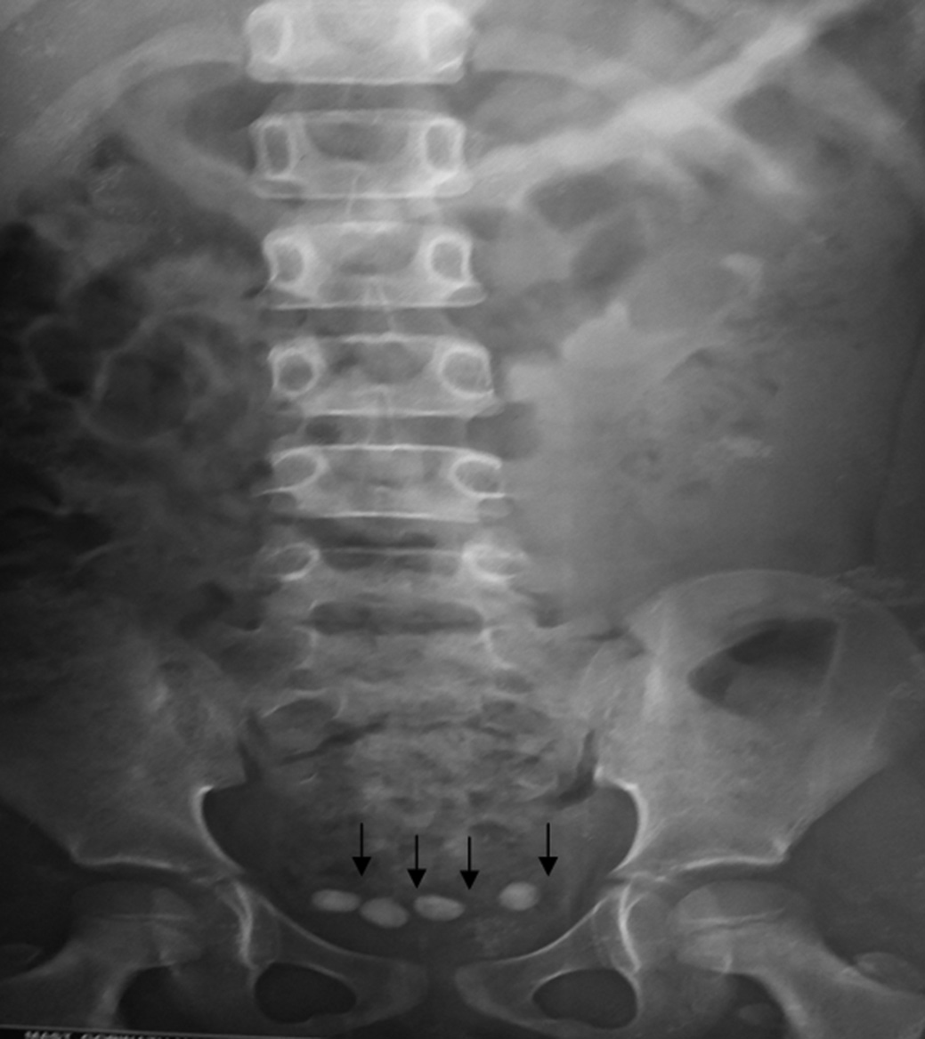
A nine-year-old male child presented in the emergency room with acute retention of urine. The catheterization through urethra was tried but failed due to obstructive bulbar urethral calculus, so suprapubic catheterization (SPC) was done. Then, the patient was referred to us. There was also the history of obstructive lower urinary tract symptoms and intermittent abdominal pain for the past two months. There was no history of any previous passage of calculus or any surgery in the past. General examination was normal. The local examination was also normal except a stone of about 8 mm was palpable over perineum at bulbar urethral region and SPC was in situ. X-ray kidney, ureter and bladder showed four radio-opaque shadows in the pelvic region [Table/Fig-1]. Intravenous urethrogram study revealed bilateral single system orthotopic ureterocele with one calculus in left-sided and two calculi in right-sided ureterocele and one calculus in the urinary bladder. The size of all the four calculi are about 7-8 mm. Both kidneys were normally excreting, there were significant bilateral hydronephrotic changes with considerable stasis of the contrast in both the ureter [Table/Fig-2] (both imaging were done before retention of urine).
Plain X-Ray KUB showed 4 calculi in pelvic region (marked with black arrows).
IVU study after 30 and 45 minutes of contrast showed bilateral ureterocele containing two calculi on right side (marked with white arrows) and one calculus on left side (marked with diamond) and one calculus in urinary bladder lumen (marked with black arrow). Normal excreting both kidneys with bilateral hydroureteronephrosis was present.

Ureteroscopy with 11 Fr pediatric cystoscope revealed a calculus of about 8 mm at mid-bulbar urethra which was pushed back to the bladder with manipulations. Cystoscopy showed bilateral orthotopic ureterocele. An adequate transurethral incision was made at the base of both ureteroceles with Bugbee electrode. Then, endoscopic extraction of two right-sided and one left-side calculi into the bladder was done with the help of grasping forceps. After that serial dilatation of earlier SPC track, a 30 Fr amplatz sheath was placed within the bladder. All the four calculi in bladder were removed with the help of 26 Fr nephroscope through the amplatz sheath. SPC was replaced and no per-urethral catheter was given. On clamping the SPC, the patient passed the urine with a good stream. SPC was removed on postoperative day 1. He was doing well on follow-up of 2 years. Follow-up USG showed gradual amelioration of hydro changes and voiding cystourethrography (VCUG) at 6, 12, and 24 months did not show any reflux.
Discussion
The ureterocele is an abnormal cystic dilatation of distal intramural end of the ureter into the bladder [1]. The most common accepted theory for its pathogenesis is the incomplete dissolution of Chwalla’s membrane [2]. An ureterocele occurs in about one in 4000 children and most common in the Caucasian population [3]. It is diagnosed more commonly in prenatal or paediatric age but occasionally can present in adult too. Female are 4-7 times more commonly affected than male. An incidence of bilateral ureterocele is only 10% [4]. Ureterocele can be classified into either single system (having single kidney, pelvicalyceal system, and ureter) or duplex system ureterocele (in which completely duplicated ureters); either orthotopic/intravesical (when the whole ureterocele lies inside bladder) or ectopic (when ureterocele fully or partially at or beyond bladder neck) [3,5]. Duplex system (up to 80%) and ectopic (approximately 80%) type are more common [6]. The present case had bilateral, orthotopic and single system ureterocele in a male child. This combination as a whole is an uncommon occurrence.
An ureterocele may be asymptomatic or may present with symptoms of urinary tract infection, renal dysfunction, and stone formation due to stasis of urine because of obstructed nature of ureterocele or its anatomical location [3]. It can also present with obstructed voiding symptom due to obstruction at bladder neck which occurs most commonly with ectopic type. Unilateral calculus in ureterocele is not uncommon (4-39%) [4]; but to the best of our knowledge, only 11 cases of bilateral orthotopic ureterocele with calculus have been reported in the literature [Table/Fig-3] [3,4,7-10]. Among these cases, no case had presented as our case with acute retention of urine due to impaction of bladder calculus at the bulbar urethra.
Review of similar reported cases in the literature
| S.No. | Authors, year | Age (in years) | Sex* | Presentation | Treatment | Follow-up | Remark |
|---|
| 1 | Singh I, 2007 [8] | 30, 38 | F,F | Flank pain, dysuria | Transverse meatotomy | 6 months | No reflux |
| 2 | Mizuno K et al., 2008 [9] | 38 | F | Pollikisuria | Endoscopic Incision | 8 months | No reflux |
| 3 | Redu VD et al., 2009 [10] | 64 | F | Hematuria | Endoscopic incision | - | - |
| 4 | Shamsa A et al., 2010 [4] | 50 | M | Dysuria | Endoscopic incision | 2 months | No reflux |
| 5 | Tomaszewski et al., 2011 [7] | 25 | M | Abdominal pain, dysuria | Endoscopic incision | 3 months | No reflux |
| 6 | Adisiyun et al., 2015 [3] | 11 | M | Abdominal pain, fever | Endoscopic incision | - | - |
| 7 | The present case | 9 | M | Acute retention of urine | Endoscopic incision | 24 months | No reflux |
M-male, F-female
Although there is a risk of reflux, endoscopic incision of ureterocele is a safe, minimally invasive, effective and standard approach [4]. Singh reported two patients with bilateral orthotopic ureterocele calculus treated with endoscopic transverse meatotomy and stone removed endoscopically [8]. Mizuno et al. had also reported a similar case which was treated with bilateral endoscopic incision with transurethral removal of stone and VCUG was nonrefluxing at 8 months follow-up [9]. We have also treated our patient with a similar approach, but calculi were removed through previous SPC site and his VCUG is non-refluxing at 2 years of follow-up.
Conclusion
The bilateral ureterocele may present as urological emergency and may lead to renal failure when not been timely diagnosed and treated. The endoscopic incision is a safe and effective approach for its treatment. Close long-term follow-up is required because of the risk of vesicoureteric junction obstruction and reflux.
*M-male, F-female
[1]. Chowdhary SK, Kandpal DK, Sibal A, Srivastava RN, Management of complicated ureteroceles: Different modalities of treatment and long-term outcomeJ Indian Assoc Pediatr Surg 2014 19(3):156-61. [Google Scholar]
[2]. Stephens FD, Smith ED, Hutson JM, Coplen D, Congenital Anomalies of the Urinary and Genital Tracts 1996 OxfordIsis Medical Media:260 [Google Scholar]
[3]. Adesiyun OM, Oyinloye OI, Akande HJ, Atobatele MO, Adeniyi WA, Abdur-Rahman LO, Bilateral giant orthotopic ureterocele appearing as kissing cobra in a Nigerian childWest Afr J Radiol 2015 22(1):42-4. [Google Scholar]
[4]. Shamsa A, Asadpour AA, Abolbashari M, Hariri MK, Bilateral simple orthotopic ureteroceles with bilateral stones in an adult: A case report and review of literatureUrol J 2010 7(3):209-11. [Google Scholar]
[5]. Tahboub A, Herskovits M, Loberant N, Bilateral orthotopic ureteroceleImaging Sci Today 2010 :321 [Google Scholar]
[6]. Stunell H, Barrett S, Campbell N, Colhoun E, Torreggiani WC, Prolapsed bilateral ureteroceles leading to intermittent outflow obstructionJBR-BTR 2010 93(6):312-3. [Google Scholar]
[7]. Tomaszewski JJ, Turner RM, 2ndOst MC, Stone cobras: adult bilateral single system ureteroceles presenting with multiple calculiUrology 2011 78(4):782-3. [Google Scholar]
[8]. Singh I, Adult bilateral non-obstructing orthotopic ureteroceles with multiple calculi: endoscopic management with review of literatureInt Urol Nephrol 2007 39(1):71-4. [Google Scholar]
[9]. Mizuno K, Kamisawa H, Hamamoto S, Okamura T, Kohri K, Bilateral single-system ureteroceles with multiple calculi in an adult womanUrology 2008 72(2):294-5. [Google Scholar]
[10]. Radu VD, Bercea B, Bilateral ureteroceles with bilateral secondary hydronephrosis and pelvic ureteral calculi. Case reportRev Med Chir Soc Med Nat Iasi 2009 113(4):1151-4. [Google Scholar]