Introduction
Hermetic sealing of the root canal is the most desirable outcome of any root canal treatment, but almost always the filling of the root canal is defective, which is a multifactorial outcome. One such factor majorly influencing the obturation is the root canal sealer used.
Aim
The present study was done for evaluating microleakage in different root canal sealers.
Materials and Methods
Sixty extracted human single rooted teeeth were used in this in-vitro study. Sealers tested for microleakage in this study were zinc oxide eugenol based sealer, Sealapex, AH Plus, MTA Plus, EndoRez, Endosequence BC. All the specimens were examined under stereomicroscope for microleakage and the obtained data were statistically analysed using One-way ANOVA test and Tukey’s multiple comparision tests using the software GraphPad Prism 7.02.
Results
The Endosequence BC group showed the least dye leakage and the highest leakage was seen in Zinc oxide Eugenol based sealer.
Conclusion
Bio ceramic salers being hydrophilic show better sealing ability compared to resin based and eugenol based sealers.
Introduction
The endodontic therapy consists of elimination of bacterial load in the pulpal canal and filling of the entire root canal system three dimensionally. The gutta-percha with the sealer should provide seal both apically and laterally, thus preventing contamination of the root canal [1,2].
Incomplete obturation of the root canal accounts for 58% of endodontic failures [3,4]. The incomplete obturation may be because of incomplete instrumentation or improper obturation technique [5]. The sealers used should fill the discrepancies between the canal wall and the gutta-percha; act as a lubricant and aid in seating the gutta-percha cones. The sealers should also fill the patent accessory and lateral canals, entomb the bacteria present within the dentinal tubule and allow for the repair of the periapical tissue [6,7].
Recently, there has been improvement in the formulation of root canal sealers. The traditional zinc oxide eugenol sealers have been replaced with resin-based, silicone based, MTA based and bioceramic based sealers. In particular, bioceramic-based sealers are gaining popularity because of their alkaline pH, chemical stability within the biological environment, lack of shrinkage and are more biocompatible [8,9].
Calcium hydroxide based sealers have been introduced to have both antibacterial action and to stimulate a sterile biological closure of apical region [10]. The limitation of these sealers is that on contact with moisture the setting time is decreased which would result in poor adaptation to canal walls [6].
MTA fillapex, MTA Obtura, Endo-CPM sealer and ProRoot Endo sealer are the most commonly available MTA based sealers. These sealers are reported to have good biocompatibility and sealing properties equivalent to epoxy-based root canal sealer or the pulp canal sealer. When in contact with simulated body fluids, MTA based sealers release calcium and encourage the deposition of apatite crystals [10,11].
EndoREZ (Ultradent Products Inc, South Jordan, UT) is a dual-cured radiopaque urethane dimethacrylate based sealer that is
used with resin coated gutta-percha. This system does not employ dentin adhesives and relies on penetration of hydrophilic resins into the dentin tubules after removal of smear layer. Recently, the bond strength and apical seal of the endorez sealer was improved using dual cured self-etching primer/adhesives [12]. The Endorez system has been shown to have satisfactory sealing ability and an easy delivery system [13].
Endosequence BC sealer and iRootSP root canal sealer are bioceramic based root canal sealers which are available as a pre-mixed white hydraulic cement paste and usually contain calcium silicate and/or calcium phosphate [14].
The most common technique for evaluating the sealing ability of the root canal sealer is the dye penetration technique which is based on the linear measurement of the dye penetration between the root filling and the canal wall. The study aimed at evaluating the sealing ability of zinc-oxide eugenol, Sealapex, AH plus, EndoRez, MTA Plus and Endosequence BC sealer using stereomicroscopic analysis of dye penetration.
Materials and Methods
Six root canal sealers were tested in vitro for their sealing ability using linear dye penetration method. Sixty freshly extracted single-rooted teeth were collected from the Department of Oral and Maxillofacial Surgery, St. Joseph Dental College and Hospital, Eluru, Andhra Pradesh, India between October 2015 to March 2016. The extracted teeth with evidence of resorption, craze lines, severe curvatures and/or incomplete apex formation were excluded from the samples. The extracted teeth were cleaned of visible debris and calculus ultrasonically and placed in 2.5% hypochlorite for two hours and stored in normal saline until further use.
The crown of all the teeth were sectioned to obtain a standardized length of 15 mm. Access cavity was prepared with Endo access bur (Dentsply, malliffer). On entering the pulp chamber, #10k file was introduced into canal to check for the canal patency. The working length was determined with #15 k file inserted into the canal until
the instrument tip was visible from the apex. The final working length was obtained after shortening 1 mm from the real root canal length.
Biomechanical preparation was carried with crown-down approach using manual Mani k-files (MN, Mani). The coronal part of the canal was enlarged with gates glidden drills 3, 2, 1 used sequentially. Next, the apical part of the canal was enlarged up to the ISO size of 40 k file. At each change of file, canals were irrigated with 1.0 mL of 3% NaOCl. After instrumentation was completed, 3.0 mL of 17% EDTA were introduced and allowed to remain in canal for five minutes [15]. Next, a final flush with 1 ml of NaOCl followed by 5.0 mL of saline was performed.
Grouping of the Samples
The 60 samples were randomly allocated to one of the group, each group consisting of 10 samples each. Group I- zinc oxide eugenol (ZOE) based sealer (Prime-Dent), Group II-Sealapex (SybronEndo), Group III-AH Plus (Dentsply), Group IV- MTA Plus (PREVEST DenPro), Group V- EndoRez (ULTRADENT PRODUCTS.INC.), Group VI- Endosequence BC (BRASSELER USA). The composition of each sealer is mentioned in [Table/Fig-1].
Composition of root canal sealers used In this study.
| Sealer | Composition |
|---|
| ZOE (Prime-Dent) | Powder: Zinc oxide, white rosin, zinc stearate, zinc acetate, magnesium oxide Liquid: Eugenol, olive oil |
| SEALAPEX (SybronEndo) | Base: Calcium oxide, bismuth trioxide, zinc oxide, sub-micron silica, zinc stearate, titanium dioxide, tricalcium phosphate Catalyst: Blend, ethyl toluene sulfonamide, poly (methylene methyl salicylate) resin, isobutyl salicylate |
| AH PLUS (DENTSPLY) | Epoxide paste: Diepoxide, calcium tungstate, zirconium oxide, aerosil, pigment Amine paste: 1-adamantane amine N,N’-dibenzyl-5-oxa-nonandiamine-1,9, TCD-Diamine, calcium tungstate, zirconium oxide, aerosil, silicone oil |
| MTA PLUS (PREVEST DenPro) | Powder: Mixture of calcium oxide, silicon oxide, bismuth oxide Gel: Hydrated polymer gel |
| EndoRez (ULTADENT PRODUCTS.INC.) | A urethane dimethacrylate resin-based endodontic sealer |
| Endosequence BC (BRASSELER USA) | Zirconium oxide, calcium silicates, calcium phosphate monobasic, calcium hydroxide, filler and thickening agents. |
Obturation of Root Canals
Obturation of the root canals was performed using lateral compaction technique. Briefly, the sealers were manipulated according to the manufacturer’s instructions. The sealers were coated to the canal wall using lentulospirals. The master cone selected was coated with sealer and passively placed to the working length. The spreader was placed beside the gutta-percha to create sufficient space for the accessory cones. The obturation was continued till the spreader no longer penetrates beyond the CEJ. The protruding gutta-percha was seared at the orifice and vertical compaction of the gutta-percha was performed. The access cavity was sealed with glass-ionomer cement.
Preparation of Specimen for Stereomicroscopic Analysis of Dye Penetration
The samples were dried for two minutes and two layers of nail varnish was applied on the external root surfaces except at the apical 2 mm to allow dye penetration into the accessory and lateral canals. The samples were immersed in freshly prepared 1% methylene blue dye for 72 hours. After three days, the nail varnish was scrapped off from the root surface, the roots were longitudinally sectioned for stereomicroscopic imaging of the vertical dye penetration at 30X magnification. Microleakage associated with different root canal sealers was evaluated and the measurement of dye penetration obtained in micrometers.
Scoring of the Vertical Dye Penetration
For the convenience of explaining the vertical dye penetration, the samples were scored as follows: Score 1- 1-3 mm; Score 2- 3-5 mm; and Score 3- >5 mm.
Statistical Analysis
The results were analysed using One-way ANOVA analysis and Tukey’s multiple comparison tests in Graph pad prism software at p<0.05.
Results
Sixty percent samples of Group VI had a score of 1 and the rest score 2. Ninety percent of the ZOE sealer group, 70% of AH plus sealer group had scores of 3. All the samples of Sealapex and MTA Plus had scores of 2 and 90% of EndoRez samples had scores of 2 [Table/Fig-2].
Distribution of all samples as per the dye leakage scoring criteria.
| Groups | Score 1 | Score 2 | Score 3 |
|---|
| Zinc oxide eugenol sealer | 0 | 1 | 9 |
| Sealapex | 0 | 10 | 0 |
| AH Plus | 0 | 3 | 7 |
| MTA Plus | 0 | 10 | 0 |
| EndoRez | 1 | 9 | 0 |
| Endosequence BC | 6 | 4 | 0 |
Means and standard deviations of the tested groups are represented in [Table/Fig-3]. The highest mean dye penetration was seen in Group I (ZOE), whereas the least microleakage was seen in Group VI (Endosequence BC). Also statistical significance (p<0.05) was seen in comparison between most of the groups except for Group I vs Group III and Group II vs Group IV [Table/Fig-4]. Stereomicroscopic images showing linear dye penetration are shown in [Table/Fig-5].
One-way ANOVA analysis showing mean and Standard Deviation (SD) for all groups.
| One-way anova analysis | group 1 | group II | group III | group IV | group V | group VI |
|---|
| F | 84.84 | | | | | | |
| p | <0.001 | | | | | | |
| Mean | | 5.476 | 4.252 | 5.319 | 4.436 | 3.379 | 2.916 |
| SD | | 0.314438 | 0.30206 | 0.583485 | 0.276976 | 0.302488 | 0.193689 |
Tukey’s multiple comparison between all the groups.
| Tukey’s multiple comparisons test | Mean Diff. | 95.00% Ci of diff. | p-value |
|---|
| Group I vs. Group II | 1.224 | 0.7611 to 1.687 | <0.001**** |
| Group I vs. Group III | 0.157 | -0.3059 to 0.6199 | 0.9152 |
| Group I vs. Group IV | 1.04 | 0.5771 to 1.503 | <0.001**** |
| Group I vs. Group V | 2.097 | 1.634 to 2.56 | <0.001**** |
| Group I vs. Group VI | 2.56 | 2.097 to 3.023 | <0.001**** |
| Group II vs. Group III | -1.067 | -1.53 to -0.6041 | <0.001**** |
| Group II vs. Group IV | -0.184 | -0.6469 to 0.2789 | 0.8469 |
| Group II vs. Group V | 0.873 | 0.4101 to 1.336 | <0.001**** |
| Group II vs. Group VI | 1.336 | 0.8731 to 1.799 | <0.001**** |
| Group III vs. Group IV | 0.883 | 0.4201 to 1.346 | <0.001**** |
| Group III vs. Group V | 1.94 | 1.477 to 2.403 | <0.001**** |
| Group III vs. Group VI | 2.403 | 1.94 to 2.866 | <0.001**** |
| Group IV vs. Group V | 1.057 | 0.5941 to 1.52 | <0.001**** |
| Group IV vs. Group VI | 1.52 | 1.057 to 1.983 | <0.001**** |
| Group V vs. Group VI | 0.463 | 0.0001434 to 0.9259 | 0.0499* |
Denotes the significant values.
denotes the highly significant values.
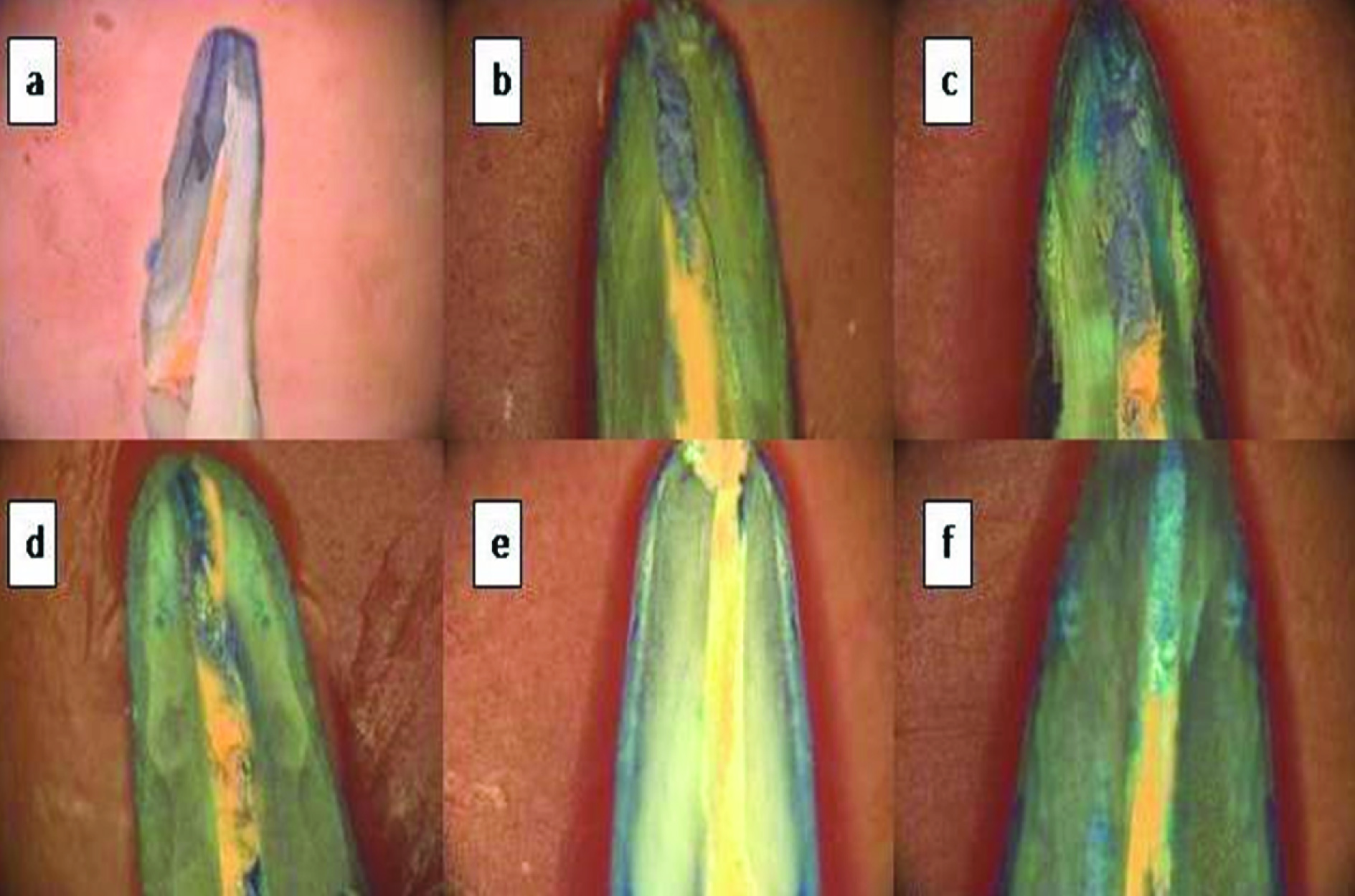
Stereomicroscopic images of tested samples showing linear dye penetration: a) Endosequence BC; b) Sealapex; c) Zinc oxide eugenol; d) AH Plus; e) Endorez; f) MTA Plus.
Discussion
The sealing of the root canal apically by the sealer is important to prevent communication of root canal contents with periapical tissue. The properties of sealers like flow, consistency, setting characteristics, solubility and adhesion to root canals are important in obtaining a hermetic seal of the root canal [16]. Though, hermetic seal is not always attainable by the currently used sealers, fluid tight seal is atleast desirable. The antimicrobial property of the sealers play a role in providing the sealing by limiting the proliferation of bacteria within the root canal and preventing the development of periapical pathology [17]. The incomplete obturation of root canal with poor apical sealing has been one of the major causes of endodontic failures. So this study was done to evaluate the sealing ability of six different sealers and hence both traditional and newer sealers were compared.
Lateral compaction technique of gutta-percha was used in this study as it has been used as a standard control for comparison [5]. Many studies have found no significant differences in the filling technique used [18-20]. Different methods are used to evaluate the apical sealing ability of root canal sealers. Linear measurement of dye penetration is the one such method which is most common, relatively easy and fast to gauge the microleakage of the sealers [21]. Different dyes are used like Methylene Blue (MB), India ink, eosin, Procion, brilliant blue, 50% silver nitrate and pelican ink. Of all, the Methylene Blue is widely used dye and the concentrations of MB used are 0.25, 1 and 2%. In our study, we used 1% MB as it was most commonly used concentration [22,23]. Ahlberg KM et al., studied the linear leakage patterns of two dyes, MB and India ink [24]. It was concluded that MB is superior in terms of penetration and has low molecular weight similar to that of bacterial toxins.
Endosequence BC sealer is a premixed calcium phosphate silicate based sealer dispersed in non-aqueous but water-miscible carriers which hardens on exposure to moisture in root dentin. In our study, Endosequence BC sealer demonstrated lower leakage values compared to all other sealers. Hegde V and Arora S in a study demonstrated lower apical leakage values in the hydrophilic sealers, similarly Pawar SS et al., in his study demonstrated lower vertical dye penetration values in Endosequence BC and Real Seal SE sealers. Also, Ersahan S and Aydin C in his study demonstrated lower apical leakage values in iRootSP (hydrophilic sealer) and AH Plus, results of these studies are in agreement with our study [25-27]. Candeiro GT et al., demonstrated acceptable flow values which correlated with ISO 6786/2001 recommendations [8]. Though, the solubility of Endosequence BC sealer was comparable to the other sealers tested it didn’t show any influence on the dimensional stability and sealing ability of this sealer [15]. Al-Haddad A et al., demonstrated marginal gaps in the Endosequence BC sealer group when the canal was irrigated with EDTA [28]. EDTA has been shown to decrease the wetting ability of sealer to dentin.
Endorez is an UDMA resin based sealer which has been shown to have hydrophilic characteristics and easy delivery system [12,13]. The Endorez sealer showed significantly less leakage compared to AH Plus sealer. The hydrophilic nature of the sealer along with ability of Endorez to form long resin tags with thin hybrid layer may account for their superior sealing ability [29]. On other hand, AH Plus sealer does not bond to gutta-percha and has been shown to lack hybrid layer. The disadvantage noted was that there may be significant amount of polymerization shrinkage when sealer and gutta-percha was passively placed without compaction. Gillespie et al., showed that the sealing ability of Endorez can be improved by using adhesive-modified Endorez filling technique [31]. De-Deus G et al., reported less bacterial leakage at nine weeks for Endorez when used in thin layers compared with AH Plus and Sealapex, but the same was not seen when used in thicker layers [32]. Our result is in agreement with Zmener O et al., who demonstrated better apical sealing when sealer was used on moist root canal dentin [32]. Sevimay S and Kalayci A found AH Plus to have better sealing ability than Endorez [13]. SEM examination in their study revealed poor penetration and adaptation of sealer to dentin in apical third for Endorez compared to AH Plus. Adanir N et al., found no significant difference in apical sealing of Endorez, AH 26 and Diaket [1]. When long term sealing ability was studied, AH Plus has shown less leakage. This may be attributed to its consistent continuous expansion of 1.2% [33-35]. In our study, we have used second generation Endorez which would have provided better result though we have used non resin coated gutta-percha.
Sealapex is a paste-paste sealer which contains calcium hydroxide in a polymeric matrix. Sealapex sealer is formulated to promote rapid healing and hard tissue formation [9]. In this study, sealapex showed less leakage compared to AH Plus, MTA plus and ZOE sealers though it was not statistically significant with MTA Plus sealer. It has been shown that the sealer being porous permits marked ingress of water and thus promoting continued reaction between powder and binder. This continued reaction during setting under humidity results in volumetric expansion and may result in decreased leakage. Cobankara FK et al., studied the apical seal of four sealers using computerized fluid filtration method. Sealapex demonstrated significantly less leakage compared to AH Plus, polymeric based and ZOE based sealers. Few other studies found no significant differences in apical leakage between sealpex and AH Plus [37].
MTA based sealers have been reported to deposit calcium phosphates in form of apatite and carbonated apatite when in contact with simulated body fluid [10,38]. These materials had similar sealing properties to epoxy based sealer [39] and pulp canal sealer [10] when evaluated using the fluid filtration system. Though, these MTA based sealers have excellent biological properties, the sealer does not bond to either dentin or gutta-percha [40,41] which may be the reason for increased leakage. Because of the short working time, MTA Plus sealer resulted in improper coating of canal during subsequent lateral compaction.
In our study, ZOE based sealers exhibited highest dye penetration compared to other sealers which is in agreement with other studies [2,20,30,31,42]. The ZOE based sealers have shown to have poor sealing and adhesion properties to dentin. Moreover, in aqueous environment, the solubility is more resulting in dissociation of zinc eugenolate in to zinc hydroxide and eugenol [43]. It was shown that when ZOE based sealer was placed in a totally dried canal, the leakage exhibited by the sealer was less [44].
Limitation
This is an invasive study in which the samples were sectioned longitudinally and during the sectioning there is a chance of gutta percha being withdrawn from the samples altering the results of this study.
Conclusion
Sealing of the canal with the use of gutta percha core and the sealer remains the main stay for the obturation of the root canal. Traditional, zinc oxide eugenol based sealers have been surpassed by the Bioceramic based sealers which have better sealing ability, chemical bonding with dentin, ease of placement and osseoconductive property all of which enhances the sealing of the root canal. With these favourable properties, the Endosequence BC sealer can be recommended as a sealer for endodontic grafting.
*Denotes the significant values.
****denotes the highly significant values.
[1]. Adanir N, Cobankara FK, Belli S, Sealing properties of different resin-based root canal sealersJ Biomed Mater Res B Appl Biomater 2006 77:01-04. [Google Scholar]
[2]. Dultra F, Barroso JM, Carrasco LD, Capelli A, Guerisoli DM, Pécora JD, Evaluation of apical microleakage of teeth sealed with four different root canal sealersJ Appl Oral Sci 2006 14:341-45. [Google Scholar]
[3]. Ingle JI, Taintor JF, Textbook of endodontics5th ed [Google Scholar]
[4]. Grossman Li, Oliet S, 1998 11th edDelrio CEEndodontic practice [Google Scholar]
[5]. Wu MK, van der Sluis LW, Ardila CN, Wesselink PR, Fluid movement along the coronal two-thirds of root fillings placed by three different gutta-percha techniquesInt Endod J 2003 36:533-40. [Google Scholar]
[6]. Ehsani M, Dehghani A, Abesi F, Khafri S, Dehkordi SG, Evaluation of apical micro-leakage of different endodontic sealers in the presence and absence of moistureJ Dent Res Dent Clin Dent Prospects 2014 8:125-29. [Google Scholar]
[7]. Loushine BA, Bryan TE, Looney SW, Gillen BM, Loushine RJ, Weller RN, Setting properties and cytotoxicity evaluation of a premixed Bioceramic root canal sealerJ Endod 2011 37:673-77. [Google Scholar]
[8]. Candeiro GT, Correia FC, Duarte MA, Ribeiro-Siqueira DC, Gavini G, Evaluation of radiopacity, ph, release of calcium ions, and flow of a bioceramic root canal sealerJ Endod 2012 38:842-45. [Google Scholar]
[9]. Desai S, Chandler N, Calcium hydroxide-based root canal sealers:a reviewJ Endod 2009 35:475-80. [Google Scholar]
[10]. Camilleri J, Gandolfi MG, Siboni F, Prati C, Dynamic sealing ability of MTA root canal sealerInt Endod J 2011 44:9-20. [Google Scholar]
[11]. Weller RN, Tay KC, Garrett LV, Mai S, Primus CM, Gutmann JL, Microscopic appearance and apical seal of root canals filled with gutta-percha and ProRoot Endo Sealer after immersion in a phosphate-containing fluidInt Endod J 2008 41:977-86. [Google Scholar]
[12]. Tay FR, Pashley DH, Monoblocks in root canals - a hypothetical or a tangible goalJ Endod 2007 33:391-98. [Google Scholar]
[13]. Sevimay S, Kalayci A, Evaluation of apical sealing ability and adaptation to dentine of two resin-based sealersJ Oral Rehabil 2005 32:105-10. [Google Scholar]
[14]. Kossev D, Stefanov V, Ceramics-based sealers as new alternative to currently used endodontic sealersRoots 2009 1:42-48. [Google Scholar]
[15]. Torabinejad M, Khademi AA, Babagoli J, Cho Y, Johnson WB, Bozhilov K, A new solution for the removal of the smear layerJ Endod 2003 29:170-75. [Google Scholar]
[16]. Zhou HM, Shen Y, Zheng W, Li L, Zheng YF, Haapasalo M, Physical properties of 5 root canal sealersJ Endod 2013 39:1281-86. [Google Scholar]
[17]. Gnana Seelan R, Arvind Kumar A, Jonathan Emil Sam R, Uma Maheswari S, Antimicrobial efficacy of different root canal sealers by using real-time polymerase chain reaction:An ex vivo studyJ Conserv Dent 2015 18:474-78. [Google Scholar]
[18]. Estrela C, MamedeNeto I, Lopes HP, Estrela CR, Pécora JD, Root canal filling with calcium hydroxide using different techniquesBraz Dent J 2002 13:53-56. [Google Scholar]
[19]. Ok E, Ertas H, Saygili G, Gok T, Effect of photo-activated disinfection on bond strength of three different root canal sealersEur J Dent 2014 8:85-89. [Google Scholar]
[20]. Kocak MM, Er O, Saglam BC, Yaman S, Apical leakage of epiphany root canal sealer combined with different master conesEur J Dent 2008 2:91-95. [Google Scholar]
[21]. Roggendorf MJ, Ebert J, Petschelt A, Frankenberger R, Influence of moisture on the apical seal of root canal fillings with five different types of sealerJ Endod 2007 33:31-33. [Google Scholar]
[22]. Baker PS, Oguntebi BR, Effect of apical resections and reverse fillings on Thermafil root canal obturationsJ Endod 1990 16:227-29. [Google Scholar]
[23]. Jacobsen EL, Shugars KA, Sealing efficacy of a zinc oxide-eugenol cement, a cyanoacrylate, and a cavity varnish used as root canal cementsJ Endod 1990 16:516-19. [Google Scholar]
[24]. Ahlberg KM, Assavanop P, Tay WM, A comparision of apical dye penetration patterns shown my methylene blue and india ink in root filled teethInt Endod J 1995 28:30-34. [Google Scholar]
[25]. Hegde V, Arora S, Sealing ability of three hydrophilic single-cone obturation sys tems:An in vitro glucose leakage studyContemp Clin Dent 2015 6:S86-89. [Google Scholar]
[26]. Pawar SS, Pujar MA, Makandar SD, Evaluation of the apical sealing ability of bioceramic sealer, AH plus & epiphany:An in vitro studyJ Conserv Dent 2014 17:579-82. [Google Scholar]
[27]. Ersahan S, Aydin C, Solubility and apical sealing characteristics of a new calcium silicate-based root canal sealer in comparison to calcium hydroxide-, methacrylate resin- and epoxy resin-based sealersActa Odontol Scand 2013 71:857-62. [Google Scholar]
[28]. Al-Haddad A, Abu Kasim NH, Che Ab Aziz ZA, Interfacial adaptation and thick ness of bioceramic-based root canal sealersDent Mater J 2015 34:516-21. [Google Scholar]
[29]. Sagsen B, Er O, Kahraman Y, Orucoglu H, Evaluation of microleakage of roots filled with different techniques with a computerized fluid filtration techniqueJ Endod 2006 32:1168-70. [Google Scholar]
[30]. Tay FR, Loushine RJ, Weller RN, Kimbrough WF, Pashley DH, Mak Y, Ultrastructural evaluation of the apical seal in roots filled with a polycaprolactone-based root canal filling materialJ Endod 2005 31:514-19. [Google Scholar]
[31]. Gillespie WT, Loushine RJ, Weller RN, Mazzoni A, Doyle MD, Waller JL, Improving the performance of EndoREZ root canal sealer with a dual-cured two-step self-etch adhesive. II. Apical and coronal sealJ Endod 2006 32:771-75. [Google Scholar]
[32]. De-Deus G, Coutinho-Filho T, Reis C, Murad C, Paciornik S, Polymicrobial leakage of four root canal sealers at two different thicknessesJ Endod 2006 32:998-1001. [Google Scholar]
[33]. Zmener O, Pameijer CH, Serrano SA, Vidueira M, Macchi RL, Significance of moist root canal dentin with the use of methacrylate-based endodontic sealers:an in vitro coronal dye leakage studyJ Endod 2008 34:76-79. [Google Scholar]
[34]. Ørstavik D, Nordahl I, Tibballs JE, Dimensional change following setting of root canal sealer materialsDent Mater 2001 17:512-19. [Google Scholar]
[35]. Paqué F, Sirtes G, Apical sealing ability of Resilon/Epiphany versus gutta-percha/ AH Plus:immediate and 16-months leakageInt Endod J 2007 40:722-29. [Google Scholar]
[36]. Bouillaguet S, Shaw L, Barthelemy J, Krejci I, Wataha JC, Long-term sealing ability of pulp canal sealer, AH-Plus, GuttaFlow and EpiphanyInt Endod J 2008 41:219-26. [Google Scholar]
[37]. Cobankara FK, Orucoglu H, Sengun A, Belli S, The quantitative evaluation of apical sealing of four endodontic sealersJ Endod 2006 3:66-68. [Google Scholar]
[38]. Xu Q, Fan MW, Fan B, Cheung GSP, Hu HL, A new quantitative method using glucose for analysis of endodontic leakageOral Surg Oral Med Oral Pathol Oral Radiol Endod 2005 99:107-11. [Google Scholar]
[39]. Tay FR, Pashley DH, Rueggeberg FA, Loushine RJ, Weller RN, Calcium phosphate phase transformation produced by the interaction of the portland cement component of white mineral trioxide aggregate with a phosphate-containing fluidJ Endod 2007 33:1347-51. [Google Scholar]
[40]. Weller RN, Tay KC, Garrett LV, Mai S, Primus CM, Gutmann JL, Microscopic appearance and apical seal of root canals filled with gutta-percha and ProRoot Endo Sealer after immersion in a phosphate-containing fluidInt Endod J 2008 41:977-86. [Google Scholar]
[41]. Hatibovic-Kofman S, Raimundo L, Zheng L, Chong L, Friedman M, Andreasen JO, Fracture resistance and histological findings of immature teeth treated with mineral trioxide aggregateDent Traumatol 2008 24:272-76. [Google Scholar]
[42]. Polineni S, Bolla N, Mandava P, Vemuri S, Mallela M, Gandham VM, Marginal adaptation of newer root canal sealers to dentin:A SEM studyJ Conserv Dent 2016 19:360-63. [Google Scholar]
[43]. Gupta S, Das G, Clinical and radiographic evaluation of zinc oxide eugenol and metapex in root canal treatment of primary teethJ Indian Soc Pedod Prev Dent 2011 29:222-28. [Google Scholar]
[44]. Hosoya N, Nomura M, Yoshikubo A, Arai T, Nakamura J, Cox CF, Effect of canal drying methods on the apical sealJ Endod 2000 26:292-94. [Google Scholar]