Autogenous bone grafts are frequently used for the reconstruction of osseous defects in maxillofacial region. Iliac crest is the most common donor site for autogenous bone grafting as cortical bone, cancellous bone or combination of both can be harvested in abundance depending upon the need [1]. Ilium provides highest concentration of osteo-component cells and greater quality of bone with less morbidity [2,3].
Several surgical techniques of hip bone harvest have been developed with intent of reducing morbidity; trephination, separate lateral or medial osteo-periosteal flaps (trapdoor), combined crestal, lateral and medial myo-oseous flaps, and saggital crestal splits [4].
Technically, harvesting iliac bone is relatively simple. However, as in all surgical procedures, a number of potential complications exist. Minor complications after harvesting iliac crest bone include prolonged pain, hypersensitivity and buttock anaesthesia [5,6]. Among the major complications that have been described are meralgia paraesthetica [7], herniation [8], disturbance of gait, arterio-venous fistula [9] and uretal injury [10]. Another uncommon but significant complication of iliac autografts is postoperative adynamic [8,10] or paralytic ileus.
The purpose of this study was to evaluate prospectively, the donor site morbidity associated with autogenous iliac crest bone grafting for reconstruction in maxillofacial region.
Materials and Methods
A prospective study was conducted which included 12 patients, who had undergone iliac crest bone harvesting for various maxillofacial and reconstructive surgical procedures like cleft alveolous repair, malar augmentation, mandibular reconstruction following tumour resection and cyst enucleation and other surgical procedures performed in the Department of Oral and Maxillofacial Surgery, Kalinga Institute of Dental Sciences, Bhubaneswar, Odisha, India, over a span of two years between January 2014 and December 2015. A written informed consent was taken from each of the patients. The sample size was calculated according to the pattern of number of cases reporting to the Outpatient Department (OPD) of the hospital and thereby reporting for the follow-ups judiciously.
The study was conducted following ethical approval of the institution and in accordance to the Declaration of Helsinki.
In these patients cancellous, corticocancellous and bicortical grafts were obtained by using superior - medial or superior - lateral or combination of both the approaches. Depending upon the age of the patient and purpose of the grafting, “Trap door” and “Tschopps” approaches were used to gain access to the cancellous bone. Routine preoperative work up, which included haematological investgations like TLC, DLC, ESR, Hb%, PCV, HbsAg, HIV, bleeding and clotting time were performed. A physician’s opinion was also obtained with regard to fitness for the surgery and patients with systemic diseases were not included in our study.
All patients received intravenous prophylactic antibiotic therapy (Amoxycillin 1 gm) and a dose of corticosteroid (dexamethasone 8 mg) to minimize the surgical oedema, half an hour prior to the surgical procedure.
Technique: Intraoperatively, after establishment of endotracheal general anaesthesia, a sand bag was placed under the ipsilateral buttock. Standard operative site preparation and draping procedures were performed, leaving the anterior part of the crest and the Anterior Superior Iliac Spine (ASIS) accessible. The iliac crest was palpated and the ASIS was located. Planned incision was marked with methylene blue. For aesthetic reasons and to avoid possible irritation of the scar from tight fitting clothes, the skin was stretched in a cranio-medial direction over the iliac crest before the incision was made. The incision was started 1 cm behind the ASIS to avoid the lateral femoral cutaneous nerve and continued posteriorly, following the iliac crest till the tubercle.
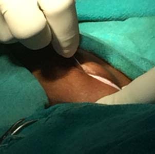
The incision was deepened through the skin, subcutaneous tissue and Scarpa’s fascia [Table/Fig-1].
Incision through skin and subcutaneous tissue.
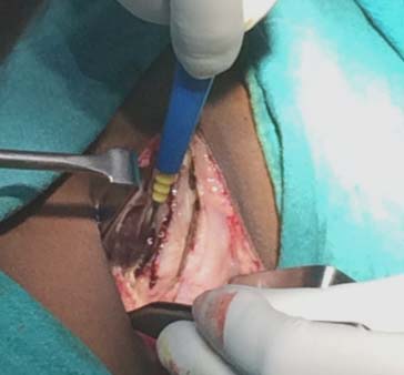
After establishing adequate haematosis, the dissection was continued sharply to the mid crest, dividing the musculo-tendenous aponeurosis of the tensor muscle of the fascia-lata and the oblique abdominal muscles, without transecting the muscle fibers. Depending upon the type of bone graft required and the age of the patients, the anterior ilium was exposed via a lateral or a medial reflection [Table/Fig-2].
Exposure of the iliac crest.
For harvesting the cancellous bone in adult, the “medial approach” and “trap door” technique were used to obtain the graft. Anterior ilium was exposed medially by stripping the medial periosteum. The “trap door” technique was initiated by making a cut on the middle third of the crest with either a stryker saw (oscillating saw) or an osteotome. Two vertical cuts on either side of the crest were made on the medial aspect. Green stick fracture was created inferiorly to gain access to the cancellous bone. The size of the trap door depends upon the amount of access desired. Once the appropriate quantity of bone was obtained with a bone gouge, the trap door was closed in its anatomical position and held in place with 3-0 vicryl suture.
In case of children for harvesting cancellous bone the “lateral approach” and “Tschopps” technique were used. The horizontal incision was placed beneath the cartilage (approximately 2 cm below the curve of the crest) and two vertical incisions upto the crest were made with a No. 15 BP blade, then the cartilagenous cap was reflected superio-medially. The cancellous bone was harvested with gauge. After obtaining adequate graft the cap was placed back in its position and its position and it was secured with 3-0 vicryl suture. Cortico-cancellous bone graft was obtained through the medial approach. The medial cortical plate was exposed directly by reflecting the iliacus muscle subperiosteally. A cortico-cancellous bone block was harvested using either oscillating saw or an osteotome. A superior cut was made midcrestally, two vertical cuts were made on either side of the superior cut, and the inferior horizontal cut was made with a curved osteotome. After removal of corticocancellous bone block from the inner table, additional cancellous bone was harvested with a gauge.
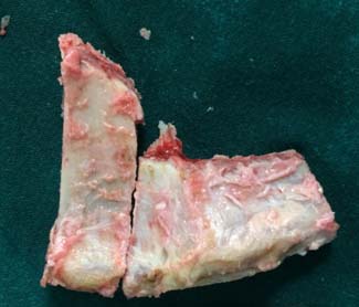
The bicortical bone grafts was harvested using both the medial and lateral approaches. Both medial and lateral cortical aspects were exposed by reflecting the muscle subperiosteally. Bicortical bone was harvested by making two vertical cuts and one inferior horizontal cut on the medial aspect with stryker saw. The cuts were deepened up to the lateral cortex. After removal of the bicortical block, the rough margins were smoothened with bone file [Table/Fig-3].
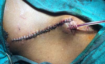
After harvesting the graft, inspection and irrigation of the wound was performed. Oozing from the marrow spaces was controlled by using electro-coagulation or bone wax. After establishment of adequate haemostasis and depending on the technique used, the pedicled cortical trap door or transected crest (cartilagenous cap) was re-approximated with resorbable sutures (3-0 vicryl). The suction drain was used depending upon the type of bone harvested, quantity and age of the patient. If it was used the suction drain was placed in the depth of the wound from a dependent stab incision. Then, the closure proceeded in layers. The periosteal layer, fascia lata and subcutaneous layers were closed using resorbable sutures (3-0 vicryl) and the skin with 3-0 silk [Table/Fig-4].
Closure of the donor site with securing of surgical drain.
Pressure dressing was then placed over the wound. All the surgeries were conducted by the same surgeon to avoid intraoperative errors.
Postoperative drugs: All patients received intravenous antibiotic (amoxycillin 1 g and metronidazole 500 mg eight hourly) for the first three postoperative days followed by oral therapy (Cap Amoxicillin 500 mg, Tab Metronidazole 400 mg eight hourly for five days. Non steroidal anti-inflammatory analgesics (Tab Diclofenac Sodium 50 mg 12 hourly) were used for first three days.
Drain removal: Drain was considered for removal, when the contents decreased to 5 to 10 ml after 48 to 72 hours.
Change of dressing: Pressure dressing was maintained and undisturbed for 48 hrs and subsequently wound dressing was changed daily till 10 days postoperatively.
Suture removal: It was considered on the ninth or 10th postoperative day after satisfactory wound healing at the site.
Evaluation of donor site: Morbidity arising from the donor site was assessed intraoperatively and postoperatively as outlined in the proforma, which also contained a detailed questionnaire for patient.
Pain at donor site: It was graded on a visual analogue scale (zero representing no pain, ten representing severe pain) to grade the subjective hip related pain at the following postoperative intervals 1, 3, 5 and 7 day, 2 weeks, 4 weeks and 8 weeks; 3 months and 6 months.
Gait: It was recorded by assessment of limp, deviation of gait, unsteadiness and unequalness of stride while walking a distance of 20 feet.
Neurosensory testing: Sensory deficit (anaesthesia, paraesthesia, dysthesia) in the femoral, gluteal and inguinal region was tested with a wisp of cotton (the subject should be able to count the number of contacts with the eyes closed).
Scar: It was assessed for its length and width with a centimeter scale.
Contour defect: It was assessed by palpation and graded as small/medium/large.
Other complications: Infections/seroma were observed and recorded in the proforma.
A simple questionnaire was framed consisting of following eight questions to evaluate the patient response on the day of discharge:
What was the duration and severity of postoperative pain at donor site?
Duration:
Severity: Nil/ Mild/ Moderate/ Severe
Was there any numbness in the hip or leg?
No numbness/ numbness in the leg/ numbness in the hip/ numbness over the scar.
Was there any problem with the wound healing of hip donor site?
No problem/ Infection/ Drainage/ Swelling
How long after the surgery did you become ambulatory?
Did you take any assistance in walking?
If yes (then for how long).
Was there any cosmetic hindrance?
Where was the pain discomfort maximum?
Donor site:
Recipient Site:
Considering the recovery experience would you be willing to undergo similar operation again?
Yes/ No
Results
In this study iliac bone grafts were harvested in twelve patients for reconstruction of various osseous defects in the maxillo-facial region. Among these patients six were females and six were males. Their age ranged from nine to 46 years with a mean age of 22.16 years.
Iliac bone was used for reconstruction of various osseous defects like malar augmentation (3 cases), mandibular reconstruction (3 cases), orbital floor reconstruction (1 case) cleft alveolus (4 cases) and reconstruction of right hypoplastic maxilla (1 case) [Table/Fig-5].
Patient age and sex distribution with type of bone harvested.
| Case No. | Age | Sex | Purpose of Grafting | Type of Graft Obtained | approach used |
|---|
| 1 | 26 | M | Malar augmentation | Cortico- cancellous | Medial |
| 2 | 45 | M | Mandibular reconstruction | Bicortical | Medial and lateral |
| 3 | 14 | F | Closure of palatal fenestration | Cancellous | Medial |
| 4 | 18 | F | Mandibular reconstruction | Bicortical | Medial and lateral |
| 5 | 15 | M | Cleft alveolus | Cortico- cancellous | Medial |
| 6 | 16 | M | Orbital floor reconstruction | Bicortical | Medial and lateral |
| 7 | 9 | F | Cleft alveolus | Cancellous | Lateral |
| 8 | 19 | F | Cleft alveolus | Cortico- Cancellous | Medial |
| 9 | 46 | M | Mandibular reconstruction | Bicortical | Medial and lateral |
| 10 | 18 | M | Malar augmentation | Cortico- Cancellous | Medial |
| 11 | 16 | F | Left maxillary alveolus reconstruction | Cortico- Cancellous | Medial |
| 12 | 24 | F | Malar augmentation | Cortico- Cancellous | Medial |
None of the patients had pre-existing gait disturbance’s scar over the iliac region, and a history of previous trauma/surgical procedure at that area. Among these twelve patients, right iliac was harvested in eleven patients and left side in one patient. All patients were ambulated on the first postoperative day with assistance and they continued to do so the third day. However, all the patients started walking without support, but with a limp from fifth to seventh day and with a mild deviation of gait. The patients walked normally at the end of two weeks without any limp or any deviation of gait or any unsteadiness, except in bicortical graft harvested cases, who had a mild gait disturbance [Table/Fig-6]. All the patients reported no cutaneous anaesthesia in the region of surgical site, but two patients showed altered sensation in gluteal region and one patient showed in the region of distribution of lateral femoral nerve of thigh, which was recovered in one to two months.
| Case No. | Time Duration |
|---|
| Days | Week | Months |
|---|
| 1 | 3 | 5 | 7 | 2 | 4 | 8 | 3 | 6 |
| 1 | P | P | A | A | A | A | A | A | A |
| 2 | P | P | p | A | A | A | A | A | A |
| 3 | P | P | P | A | A | A | A | A | A |
| 4 | P | P | P | A | A | A | A | A | A |
| 5 | P | P | P | P | A | A | A | A | A |
| 6 | P | P | P | P | A | A | A | A | A |
| 7 | P | P | A | A | A | A | A | A | A |
| 8 | P | P | P | A | A | A | A | A | A |
| 9 | P | P | P | P | A | A | A | A | A |
| 10 | P | P | P | A | A | A | A | A | A |
| 11 | P | P | A | A | A | A | A | A | A |
| 12 | p | p | P | A | A | A | A | A | A |
P = Present, A = Absent
Incision length ranged from 3-8 cm with a mean of 5.29 cm. Among these patients, medial approach was used in six patients, lateral approach in two patients and combination of both the approaches were used in four patients.
For all these cases the time required for exposure of iliac crest from time of incision placement was within 15 minutes with excellent exposure. Out of twelve patients, cancellous bone was harvested in two patients, corticocancellous in six patients and bicortical in four patients. The length of the iliac bone harvested was ranged from 2-7.5 cm, with a mean of 4.5 cm. The volume of cancellous from 2 to 5 cubic centimetre (cc) with a mean of 4 cc. The grafts were obtained by using a scalpel, oscillating saw, osteotome and a bone gouge.
None of these patients had intraoperative complications like haemorrhage, damage to the muscles/ligaments, fracture of the ilium and damage to the acetabular fossa/femur head.
Pain was quite severe in all cases on the first postoperative day, by the third postoperative day pain was moderate and by the fifth postoperative day pain became tolerable. On the seventh postoperative day, mild pain was experienced by all these patients and none of these patients complained of any pain at end of fourth week [Table/Fig-7].
Donor site pain assessment.
| Case No. | Time Duration |
|---|
| Days | Week |
|---|
| 1 | 3 | 5 | 7 | 2 | 4 | 8 |
|---|
| 1 | 8 | 5 | 2 | 1 | 0 | 0 | 0 |
| 2 | 8 | 6 | 4 | 3 | 1 | 0 | 0 |
| 3 | 9 | 8 | 5 | 4 | 1 | 0 | 0 |
| 4 | 8 | 6 | 3 | 2 | 0 | 0 | 0 |
| 5 | 8 | 5 | 3 | 2 | 0 | 0 | 0 |
| 6 | 7 | 5 | 3 | 3 | 1 | 0 | 0 |
| 7 | 7 | 5 | 4 | 2 | 0 | 0 | 0 |
| 8 | 8 | 6 | 4 | 3 | 0 | 0 | 0 |
| 9 | 8 | 7 | 5 | 3 | 2 | 0 | 0 |
| 10 | 6 | 5 | 3 | 1 | 0 | 0 | 0 |
| 11 | 7 | 5 | 4 | 2 | 1 | 0 | 0 |
| 12 | 7 | 4 | 3 | 1 | 1 | 0 | 0 |
1-3 Mild, 4-7 Moderate, 8-10 Severe
The final scar was situated almost lateral to the crest in all these cases, the average length and breadth being 5.3 cm and 3.4 mm respectively. Mild to moderate contour defect was apparent in all cases [Table/Fig-8].
| CASE NO. | SCAR |
|---|
| Length in cm | Width in mms |
|---|
| Months | Months |
|---|
| 3 Months | 6 Months | 3 Months | 6 Months |
|---|
| 1 | 5.0 | 5.0 | 2.2 | 2.5 |
| 2 | 7.0 | 7.0 | 1.2 | 2.0 |
| 3 | 5.0 | 5.0 | 2.0 | 2.2 |
| 4 | 5.2 | 5.3 | 2.5 | 2.8 |
| 5 | 5.3 | 5.3 | 2.0 | 2.2 |
| 6 | 6.0 | 6.0 | 1.2 | 1.5 |
| 7 | 3.0 | 3.0 | 1.8 | 2.0 |
| 8 | 5.3 | 5.3 | 1.5 | 1.9 |
| 9 | 8.0 | 8.0 | 2.0 | 2.4 |
| 10 | 5.0 | 5.0 | 1.5 | 1.7 |
| 11 | 5.0 | 5.0 | 1.2 | 1.2 |
| 12 | 4.5 | 4.5 | 1.0 | 1.2 |
During the postoperative course, infection was experienced in two patients and seroma in one patient within the two weeks of postoperative period, which was resolved by incision, drainage and antibiotic therapy for five days. All these patients were discharged on 10th postoperative day.
Questionnaire Results
Questionnaire results were based upon patient’s response on discharge day.
Most of the patients reported moderate pain (60%) while fewer said that their pain was severe (20%), and nearly equal number (20%) reported mild pain.
None of these patients reported no numbness in the hip/leg.
Nine patients responded that there was no problem associated with the donor site; two patients reported that there was some discharge from the operated site and one patient reported that there was swelling in the operated area.
Approximately 98% of the patients reported walking to the bathroom within the first 24 hours following surgery.
Ninety percent of patients took assistance for walking for two days, 8% of patients for the first three days and 2% of patients for the first four days.
None of these patients had mentioned about cosmetic hindrance.
Seven patients experienced much discomfort/pain in donor site than recipient, four patients experienced more discomfort in recipient site than donor site and one patient experienced equal discomfort on both sites.
Out of 12 patients only three patients stated that based on the recovery experience, he/she would not be willing to undergo a similar operation.
Discussion
Bone grafting has been used in reconstructive surgery for over 100 years. Converse JM et al., laid the foundation for the modern use of osseous, autologous grafts in facial surgery [11].
Various types of grafts available for the reconstruction of osseous defect in maxillofacial region are autogenous bone grafts, allogenic bone grafts, xenogenic bone grafts and implants. Among these grafts autogenous grafts are superior, because of their good function, form and adaptability and also new bone formation in the recipient site by osteogenesis, osteoconduciton or osteoinduction [12].
The choice of donor site is determined by several factors, including surgeon’s preference, volume of bone required, available embryologic origin of the bone and morbidity associated with the harvest. Potential donor sites that have been used for maxillofacial reconstruction include anterior and posterior iliac crest, ribs, calvarias, mandible (chin), tibia and other sites [1,3]. Among these the anterior iliac crest is the most common donor site for autogenous bone grafting as cortical bone, cancellous bone or combination of both can be harvested in abundance depending upon the need. Ilium provides highest concentration of osteocompetent cells and greater quantity of bone with less morbidity [3]. Harvesting of anterior ilium is more advantageous than posterior ilium because of its ease of harvesting, favourable quality of bone, and ability to harvest the graft simultaneously with the oral procedures and thus reduce the operating time [13,14].
Exposure of the bony anterior ilium may be via lateral or a medial reflection. Many clinicians believed that there is less morbidity when a bone is harvested from medial aspect, because it avoids the reflection of tensor fascia muscle and therefore much less gait disturbances [1,15].
Several surgical techniques of harvesting cancellous, corticocancellous and bicortical bone graft of the anterior iliac have been developed with the intent of reducing morbidity. The complications of the anterior iliac approach have been reported by many authors. Intraoperative complications include haemorrhage, damage to the muscles/ligaments, fracture of the ilium [6], damage to the acetabu-lar fossa/femur head.
In our study, twelve patients were studied to evaluate the donor site morbidity associated with iliac crest bone harvest in maxillofacial and reconstructive surgery.
In these patients the donor site morbidity was evaluated by using intraoperative and postoperative parameters.
Blood loss from the iliac crest harvest was not very significant in our study. Muscle dissection and the length of operative time are the major contributing factors to the amount of blood loss. However, meticulous attention to surgical technique and a proper plane of dissection down to the periosteum with sub periosteal stripping and preventing damage to muscles may minimize the amount of blood loss.
In our study, we did not come across any ilium fracture, while harvesting the graft. This complication can be avoided by good handling and also by using surgical saw. But this incidence was reported by only few authors [16]. Schaaf H et al., reported only one out of 75 patients with a fracture of the ilium [17].
The incidence and severity of postoperative pain from the iliac crest donor site has been commented by several authors [6,11]. In our study mostly all patients showed mild pain at the end of first week postoperatively. None of the patients had prolonged postoperative pain that is more than two weeks. Rawashdeh MA et al., reported 76% of the patients had more pain in the donor site than they had expected [18], even Clarke A et al., reported 11% of their 33 patients had pain even in the third postoperative month [19]. Matsa S et al., showed in their study almost no complaint of pain by patients at the end of one month [20]. Meticulous surgical technique and plane of dissection down to the periosteum with subperiosteal stripping and preventing damage to the muscles may minimize the post surgical pain.
Incidence of gait disturbance followed by iliac harvesting has been reported by several authors. In our follow up 98% of our patients were found to be free of limp two weeks after surgery which may attribute to preventing damage to the muscles, prevention of the iliac spine, avoiding damage to the tensor fascia lata muscle, good reapproximation of the periosteal margins and proper layered wound closure. Beirne JC et al., stated that 94% of their patients were limp free two weeks after the procedure [2]. Matsa S et al., reported around 27.7% of their patients had abnormal gait in the first month which was completely resolved by the third month of follow up [20], while Rawashdeh MA et al., showed that all their patients reached normal gait within two weeks of the surgery [18]. Matsa S et al., showed 27.7% of their patients had abnormal gait in the first month which got fully resolved by third month [20].
Damage to the sensory nerves at the hip region, while harvesting the graft has been mentioned by many authors. In our series, the incidence of altered sensation in the distribution of lateral femoral cutaneous nerve was 8.3% (out of 12 patients) and it was restored in two months. Altered sensation in the one patient could have been due to the excessive retraction of the tissues while harvesting the bicortical iliac graft. Beirne JC et al., reported the incidence of damage to this nerve in 1.3% in their study of 137 patients [2].
In our study, we had very less percentage of involvement of this nerve which could be due to placement of the incision 2 cm away from the anterior iliac spine, placing the incision lateral to the crest and also by careful retraction of tissues. In our study, we did not come across any damage to the lateral cutaneous branch of subcoastal nerve. In our study two patients showed temporary loss of sensation along the course of lateral cutaneous branch of the iliohypogastric nerve which was restored in two months. This may be due to retraction of tissues while harvesting bicortical graft. Transection of this nerve can be avoided by limiting the extension of the incision posteriorly [2].
Good re-approximation of the periosteal margins and a proper layered wound closure may minimize the width of the scar. There is general acceptance of the surgical scar despite its dimensions since it lies in an obscure area. None of the patients complained about their surgical scar, Kalk WW et al., stated that 2% of their patients had complained about the scar [3].
None of our patients had experienced delayed wound healing, may be due to maintenance of the incision lateral to the crest.
In our study, we found mild to moderate contour defect after harvesting cancellous and bicortical grafts. This may be due to good approximation of the periosteum and harvesting of adequate bone for the reconstruction of various osseous defects.
The incidence of seroma was minimal in our study that is about 9% (1 of 12 patients). According to Tayapongsak P et al., incidence of seroma was 5% (two out of 40 patients) [1]. We treated our patient by incision and drainage. This complication could be avoided by complete haematosis of the oozing blood from the marrow surface with a thin layer of bone wax before closure, good re-approximation of the periosteal margins, proper layered wound closure and also by using drain [6].
Infection is most common postoperative complication. It is illustrated by many authors. In our study two out of 12 patients (16%) experienced infection in the second week of postoperative period. It was treated by administering antibiotics for 5 days and also by regular dressing of the wound.
Herniation of the abdominal contents through the wing of ilium following removal of bone from the site for grafting is an extremely rare complication [8]. It occurs only when the full thickness of bone has been harvested. In our study, four patients had undergone bicortical harvesting but none of our patients developed this complication. This can be avoided by preventing damage to the muscles and good approximation of the tissues.
Traumatic retroperitoneal hematoma is an unusual complication of iliac crest graft procurement. The regional origins of bleeding may be any of the adjacent visceral organs, the bone, blood vessels or muscle adjacent to the retroperitoneal space. Ziccardi VB et al., reported a retroperitoneal hematoma as a complication of anterior iliac crest harvest, which was caused by the gluteal muscle tear due to lateral cortex perforation [21]. In our study, we did not encounter this problem. This complication can be avoided by achieving good haematosis before closure, by good approximation of muscles and also by using vacuum drain.
Even though the above mentioned complications have been reported by many authors, our study encountered very minimal morbidity at the donor site, following harvest of iliac crest.
Limitation
The study had a few limitations in aspect to the small sample size and also not having a control group for the comparison. We are certain that in years to come such type of researches would be carried on at high scales to give conclusive results.
Conclusion
Considering the success rate in our study we can safely conclude that the anterior iliac crest provides an adequate harvest of cancellous, corticocancellous or bicortical grafts for reconstruction of various osseous defects in the maxillofacial region with least morbidity and should be considered as a major reservoir of bone for bony reconstructive procedures.
P = Present, A = Absent
1-3 Mild, 4-7 Moderate, 8-10 Severe