Identification of the sex of the skeletal remains is a very important step in biological profiling of the Skeletal remains or a badly burnt body in establishing the identity of the individual in forensic medicine [1,2]. Apart from sex, stature, age and race can also be determined by the anthropometric study of bones [3]. Hip bone is considered as a more suitable bone in the body for identification of sex because of its marked sexual dimorphism which is a result of childbearing and locomotion in females and males [4]. Determination of sex from hip bone by the visual method is very subjective. Discriminant analysis using biometry of hip bone rules out observer bias [5]. Various researchers have evaluated different parameters for estimation of sex in hip bone. Parameters in anterior border of the hip bone for sex determination were studied by Sachdeva K et al., [6]. It was observed that minor yet statistically significant differences exist between discriminant values of hip bones in males and females in different populations [7]. Skeletal parameters of hip bone like posterior width of greater sciatic notch diameter of acetabulum; total height of hip bone, iliac breadth, pubic length were studied by Isaac B in human hip bones of the Southern Indian population [8]. The aim of the present study was to evaluate the effectiveness of anterior border parameters of the hip bone for sex determination using discriminant function analysis in Southern Indian population.
Materials and Methods
A descriptive observational study was done that included a total number of 206 adult, dry hip bones obtained from Melmaruvathur Adiparasakthi Institute of Medical Sciences and Research, Melmaruvathur, Tamil Nadu, India, between September and October, 2012 and Subbaiah Medical College, Shivamogga, Karnataka between November and December, 2016.
Damaged or deformed hip bones were excluded from the study. Out of 206 hip bones, 121 males and 85 female bones were segregated based on morphological features by the visual method using following points:
Greater sciatic notch: Female: Wider (>90 degree); Males: Narrower(<90 degree);
Ischial spine: Females: Not inverted; Males: Inverted;
Ischiopubic ramus: Females: Not everted; Males: Everted;
Obturator foramen: Females: Triangular; Males: Oval.
After determining the sex of the hip bones, the following five parameters were measured in millimetre using digital Vernier calipers:
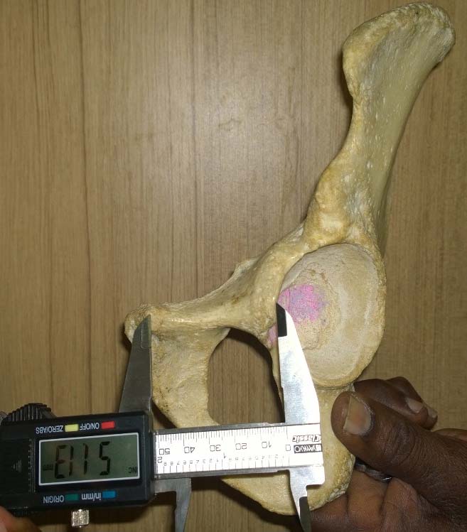
Distance between pubic tubercle to anterior rim of acetabulum [Table/Fig-1];
Distance between highest point on pubic tubercle to anterior rim of acetabulum.
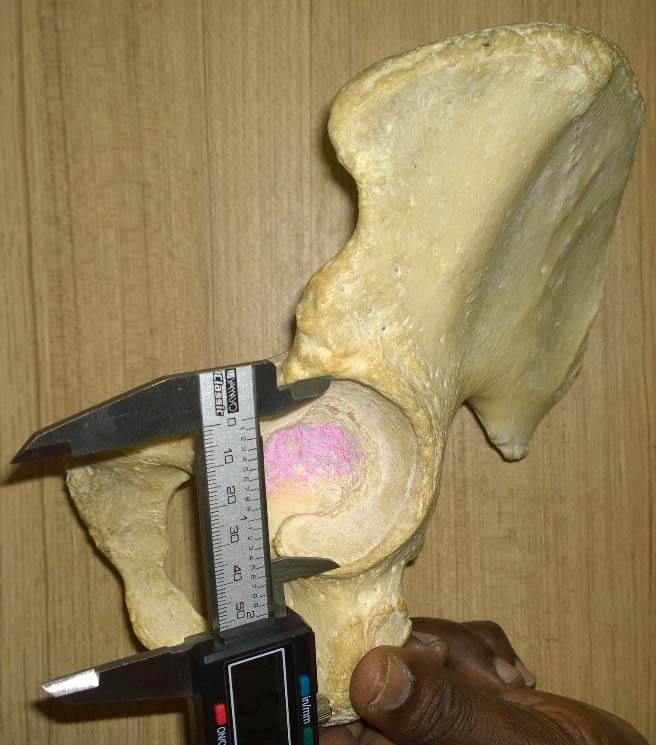
Vertical acetabular diameter [Table/Fig-2];
Vertical diameter of acetabulum. (Images left to right)
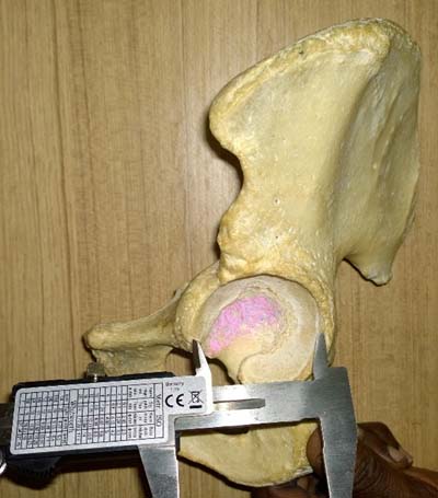
Transverse acetabular diameter [Table/Fig-3];
Transverse diameter of acetabulum.
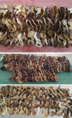
Distance between pubic tubercle to the highest point on iliopubic eminence [Table/Fig-4]; [Table/Fig-5] shows the collection of hip bones used for study.
Distance between highest point of iliopubic eminence to highest point of pubic tubercle.
(Images left to right)
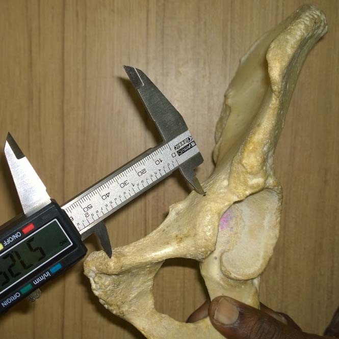
Image showing the collection of hip bones used for study.
Statistical Analysis
The prediction of sex from these hip bones was done using discriminant function analysis. Normally distributed variables are compared using Student’s t-test in Microsoft Excel 2016. The level of significance considered for this study is 5%, α=0.05.
Results
Distance between pubic tubercle to anterior rim of acetabulum (mm): The distance between pubic tubercle to anterior rim of acetabulum was greater in females than in males [Table/Fig-6].
Distance between pubic tubercle to anterior rim of acetabulum (mm).
| Details of measurements | Male | Female |
|---|
| No. of bones | 121 | 85 |
| Range (mm) | 44-65 | 49-75 |
| Mean (mm) | 54.4 | 57.85 |
| Standard deviation | 5.34 | 4.63 |
The calculated range (mean ± 3 x SD) for males was 38.38-70.42 mm and the calculated range for females was 43.96-71.74 mm. The demarking point for males was <43.96 mm and females was >70.52 mm. Below 43.93 mm, there were only males, no females; and above 70.42 mm, there were only females and no males. The limiting point between males and females was 57 mm. The sex difference between means of distance between pubic tubercle to anterior rim of the acetabulum in males and females was statistically significant with p-value: <0.001.
Vertical acetabular diameter (mm): The vertical acetabular diameter in males was greater than in females [Table/Fig-7].
Vertical acetabular diameter (mm).
| Details of measurements | Male | Female |
|---|
| No. of bones | 121 | 85 |
| Range (mm) | 40-60 | 40-57 |
| Mean (mm) | 50.85123967 | 47.22352941 |
| Standard deviation | 3.439140292 | 3.590253 |
The calculated range for males was 40.51-61.04 mm. The calculated range for females was 36.43-57.97 mm. The demarking point for males was >57.97 mm and females was <40.51 mm.
Below 40.51 mm, there were only females and no males; above 57.97 mm, there were only males and no females. The limiting point between males and females was 49.24 mm. The sex difference between means of vertical acetabular diameter in males and females was statistically significant with p-value: <0.001.
Transverse acetabular diameter (mm): The transverse acetabular diameter in males was greater than in females [Table/Fig-8].
Transverse acetabular diameter (mm).
| Details of measurements | Male | Female |
|---|
| No. of bones | 121 | 85 |
| Range (mm) | 39-56 | 37-53 |
| Mean (mm) | 48.1322314 | 45.24705882 |
| Standard deviation | 3.098231078 | 3.5183339308 |
The calculated range for males was 39.03-57.37 mm. The calculated range for females was 34.71-55.77 mm. The demarking point for males was >55.55 mm and females was <39.03 mm. Below 39.03 mm, there are only females and no males; above 55.55 mm, there only males and no females. The limiting point between males and females was 47 mm. The sex difference between means of transverse acetabular diameter in males and females was statistically significant with p-value: <0.001.
Highest point on iliopubic eminence to pubic tubercle (mm): The distance between the highest point on iliopubic eminence to pubic tubercle is greater in females than in males [Table/Fig-9].
Distance between the highest point on iliopubic eminence to pubic tubercle (mm).
| Details of measurements | Male | Female |
|---|
| No. of bones | 121 | 85 |
| Range (mm) | 40-61 | 45-66 |
| Mean (mm) | 50.8 | 52.7 |
| Standard deviation | 5.04 | 4.79 |
The calculated range for males was 35.68-65.92 mm. The calculated range for females was 38.33-67.07 mm. The demarking point for males was <38.33 mm and females was >65.92 mm. Below 38.33 mm, there are only males, no females; above 65.92 mm, there only females and no males. The limiting point between males and females was 52.12 mm. The sex difference between means of distance between highest point on iliopubic eminence to pubic tubercle in males and females was statistically significant with p-value: 0.006522.
Discussion
Sexual dimorphism is more pronounced in the hip bone among all the bones of the body, owing to its changes of the pelvis during parturition. Several nonmetric characteristic features have been associated with male hip bones like eversion of ischiopubic ramus, oval shape of obturator foramen, and shallow iliac fossa as compared to female hip bones. Earlier investigations revealed that the osteometric differences exist between different population groups [9].
Study by Sachdeva et al., employed parameters like: (i) straight distance between Anterior Superior Iliac Spine (ASIS) and Symphyseal Surface (SS) of pubis; (ii) straight distance between ASIS and Pubic Tubercle (PT); (iii) straight distance between Anterior Inferior Iliac Spine (AIIS) and SS of pubis; (iv) straight distance between AIIS and PT; (v) Arch of Anterior Interspinous notch (ASIS– AIIS); (vi) arch between AIIS and Ilio-Pubic Eminence (IP); (vii) depth of notch between AIIS and IP; (viii) arch of anterior border (ASIS–SS) for sex determination of hip bone [6].
Another study done by Dixit SG et al., had measured: (a) acetabular height (acetabular vertical diameter); (b) total pelvic height/acetabular height; (c) midpubic width/acetabular height; (d) pubic length/ acetabular height in 100 hip bones for sex determination [10].
Distance between pubic tubercle to anterior rim of acetabulum (mm): In our study, distance between pubic tubercle to anterior rim of the acetabulum, mean values were 54.4±5.3 mm and 57.8±4.6 mm in males and females respectively. Nagesh KR et al., studied the distance between the pubic symphysis and the anterior rim of acetabulum as pubic length and reported mean in males as 61.7±0.40 mm, mean in females as 64.5±0.40 mm [11]. Even though our study involved the distance between pubic tubercle to anterior rim of acetabulum as against the distance between pubic symphysis and anterior rim of acetabulum used by Nagesh KR et al., there was a similar pattern in both the studies in which higher values were observed in females with respect to pubic length, using a different landmark. However, there were no previous studies involving the pubic tubercle and anterior rim of the acetabulum. From the present study, it can be inferred that, a hip bone of unknown sex can be classified as definite male, if the measurement is <43.96 mm and a definite female, if the measurement is >70.52 mm.
Acetabulum: Many researchers had studied acetabulum and its utility towards the determination of sex in hip bones. Rajangam S et al., whose study included samples from Karnataka in India also concluded that acetabular diameter is considered as the single best parameter for determination of sex in hip bones [12]. Murphy AMC et al., reported that sex of the hip bone could be determined with 85.2% to 86.2% accuracy [13].
Vertical acetabular diameter (mm): In our study, the mean values of vertical diameter of acetabulum was 50.8±3.4 mm and 47.2±3.5 mm and p-value of <0.001 in males vs females with respect to vertical diameter of acetabulum, which is similar to the observations made by Dixit SG et al., [10]. From the present study, it can be inferred that, a hip bone of unknown sex can be classified as definite male, if the measurement is >57.97 mm and a definite female, if the measurement is <40.51 mm.
Transverse diameter of acetabulum: In a study done by Nagesh KR et al., in 67 bones, the mean transverse acetabular diameter in males was 51.1±0.26 mm and mean transverse diameter in females was 46.3±0.31 mm [11]. In our study, the mean transverse diameter in males was 48.1±3.09 mm and the mean transverse diameter in females was 45.2±3.51 mm. Our findings were similar to the pattern of findings of Nagesh KR et al., with respect to the mean in males and females. However, the standard deviation in males and females were not similar with the study of Nagesh KR et al., [11]. From the present study, it can be inferred that, a hip bone of unknown sex can be classified as definite male, if the measurement is >55.55 mm and a definite female, if the measurement is <39.03 mm.
Distance between highest point on Iliopubic eminence to pubic tubercle (mm): Gomez Pellico L et al., in his study involving multiple parameters found that the mean distance between the highest point on iliopubic eminence to pubic tubercle in males was 51.37±5.49 mm and in females was 49.48±4.93 mm [14]. In our study, the mean distance between the highest point on iliopubic eminence to pubic tubercle in males was 50.8±5.04 mm and in females was 52.7±4.79 mm and the difference in the means of males versus females was statistically significant. Our study results differ with that of Gomez Pellico L et al., with respect to the measurement of the distance between the highest point on iliopubic eminence to pubic tubercle.
From the present study, it can be inferred that a hip bone of unknown sex can be classified as male, if the distance between pubic tubercle to anterior rim of acetabulum is <43.96 mm, vertical acetabular diameter is >57.97 mm, transverse acetabular diameter is >55.55 mm, distance between highest point on Iliopubic eminence to pubic tubercle is <38.33 mm.
From the present study, it can be inferred that, a hip bone of unknown sex can be classified as definitive female, if the distance between pubic tubercle to anterior rim of acetabulum is >70.52 mm, vertical acetabular diameter is <40.51 mm, transverse acetabular diameter is <39.03 mm, distance between the highest point on iliopubic eminence to pubic tubercle is >65.92 mm.
Limitation
The present study included a sample size of 206 hip bones sourced from two Southern states of Tamil Nadu and Karnataka in India. It did not include bones from other states in Southern India or bones from other regions of India. There is a scope for more elaborate studies involving samples from different regions and different population in India to contribute to the knowledge base of forensic anatomy.
Conclusion
Identification of sex of hip bone is the first step in creating a biological profile of the skeletal remains. The present study has employed the parameters like the distance between pubic tubercle to anterior rim of the acetabulum, vertical and transverse diameters of the acetabulum, and the distance between the highest point on the Iliopubic eminence to pubic tubercle that are primarily features of the anterior part of the hip bone. The parameters used in this study will be useful for sex determination, if the conventionally used parameters like posterior part of ilium (greater sciatic notch curvature measurement), ischial tuberosity, ischiopubic ramus, ischial spine are damaged due to trauma or destroyed due to mishandling during exhumation. The parameters used in the present study can be used in conjunction with the conventionally used parameters to increase the accuracy in sex determination of hip bone.