Rickettsial diseases comprise a wide spectrum of diseases which are reported from different parts of India quiet long ago. Many cases of rickettsial diseases go undiagnos due to lack of diagnostic techniques and the reported incidence and prevalence may be an underestimation of the actual burden of the disease. A higher index of suspicion, clinical awareness and proper use of available diagnostic tools would increase the frequency of diagnosis. Gangrene is an uncommon complication in cases of rickettsial fever. Extensive gangrene of the digits or whole limb, even requiring amputation has been more commonly reported with Rocky Mountain spotted fever. These cases are being reported to highlight the occurrence of gangrene in rickettsial fever and the importance of appropriate management at the earliest.
Case 1
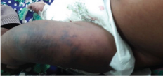
A 3-month-old female infant, presented with high grade fever for four days and rashes and vomiting for one day. She had a generalised seizure just before admission (no past history of seizures) and was in postictal state. Physical examination revealed pulse rate of 120 bpm, good volume, Capillary filling time < 3seconds, Blood Pressure (BP) 94/60 mm Hg, Respiratory Rate (RR) 38 breaths/min, temperature 99°F, pallor, pitting oedema of all the limbs along with discrete, palpable, purpuric rash [Table/Fig-1] all over the body excluding palms and soles, which appeared on the 4th day of illness.
Discrete, palpable, purpuric rashes over left leg.
There was no eschar. Abdominal examination showed haepatomegaly. Spleen was not palpable and there was no evidence of free fluid. Respiratory system examination showed crepitation in bilateral lung fields. There was no focal deficits and the rickettsial score was 15. Gangrene was developed in fingers, toes and ear lobes on second day of admission [Table/Fig-2]. She needed intubation and ventilation along with inotrope support from day 3 of admission due to respiratory distress, severe metabolic acidosis and shock, respectively.
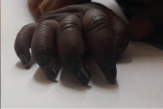
She was treated with ceftriaxone (75 mg/kg/day), in view of suspected sepsis. Azithromycin (10 mg/kg/day) was started in view of suspected ricketssial infection and given for two days. Then gangrene was gradually extended upto the middle phalanx of the three middle fingers of the right hand, so doxycycline (2.2mg/kg/ day) was added later (day 5). Mechanical ventilation was needed for three days with ionotropes. Child responded to doxycycline within 48 hours; fever subsided and progression of gangrene was stopped. She was gradually weaned off from inotrope and ventilator and was discharged in a stable condition on day 15. Surgical opinion was sought, intervention was not considered essential. There was auto-amputation of right index and middle fingers up to middle phalanx and other gangrenous areas resolved over 10-20 days with peeling of skin.
Case 2
A 2-year-old male child presented with fever, purpuric rashes involving palms and soles for 4 days duration. Rashes appeared on the 4th day of illness and there was no eschar. Physical examination revealed pulse rate of 100 bpm, normal volume, CFT<3 seconds, BP 102/68 mm Hg, RR 28 breaths/min, temperature 100°F, no hepatosplenomegaly, CNS examination was normal. Rickettsial score was 16. He developed gangrene of right toes on 2nd day of admission [Table/Fig-3].
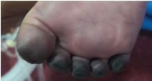
He received symptomatic treatment (antipyretics) on first day. On second day of admission, dry gangrene of the right toes were noted and started on doxycycline 4 mg/kg/day. He gradually improved, there was extensive peeling of skin at the gangrenous areas; no residual deformity and was discharged in stable condition on day 7.
Case 3
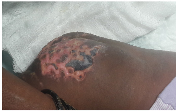
A 12-month-old female infant, presented with high grade fever for nine days and rashes and swelling of limbs for 5 days. On arrival to our hospital she had pallor, pitting oedema of all the limbs along with discrete, gangrenous areas involving knee, gluteal region and ear lobes which developed on 7th day of illness [Table/Fig-4]. Physical examination revealed pulse rate of 134 bpm, good volume, CFT< 3seconds, BP 94/56 mm Hg, RR 40 breaths/min, temperature 100°F. There was no eschar. Abdominal examination showed hepatosplenomegaly and there was no evidence of free fluid. Respiratory system examination showed crepitations in bilateral lung fields. CNS examination was normal. Rickettsial score was 19.
Gangrenous areas involving knee
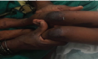
Investigations mentioned in table 6. She got admitted on the 9th day of illness with dry gangrene involving knee, gluteal region and ear lobes which developed on 7th day of illness. She improved with doxycycline (4 mg/kg/day) and the gangrenous area undergone extensive peeling leaving a raw area behind [Table/Fig-5]. There was no residual deformity and was discharged after 7days.
Investigations of all cases.
| Parameters | Normal range | Case 1 | Case 2 | Case 3 | Case 4 |
|---|
| Haemoglobin (gm/dL)Total count(/mcL)Differential countPlatelet count(/mcL)CRPUrea (mg/dL)Creatinine(mg/dL)Na(mEq/L)SGOT(iu/L)SGPT(iu/L)ALP(iu/L)S.albumin(gm/dL)Weil felix test | 11-144000-11000>P60L40150000-450000Qualitative20-400.7-1.4135-1455-407-5644-1473.5 -5.5≥1:320 | 9.912000P41L5660000Negative210.9134276723.6Positive(D6) | 10.214000P67 L32120000Negative350.613234122343.4Positive(D6) | 8.222000P60L34400000Positive200.513514161392.1Positive(D9) | 118700P 45L52340000Negative420.812938241693.8Positive(D7) |
Interpretation: Weil felix test titre of ≥1:320 was considered as diagnostic
Post treatment image of case 3 showing peling of skin with a raw area.
Case 4
A 8-year-old female child presented with fever, purpuric rashes involving palms and soles for 5 days duration. Rashes appeared on the 5th day of illness. There was no eschar. Physical examination revealed pulse rate of 96 bpm, good volume, BP 116/70 mm Hg, RR 22 breaths/min, temperature 100°F and hepatomegaly. Spleen was not palpable and there was no evidence of free fluid. CNS examination was normal. Rickettsial score was 18. She developed gangrene of left middle and index fingers on day 2 of admission.
She had dry gangrene of the tip of left middle and index fingers, noticed on day 6 of illness and started on doxycycline(4 mg/kg/day. She gradually improved with treatment and the gangrenous areas had extensive peeling and there was no residual deformities. She was discharged in a stable condition after 6 days.
Discussion
Rickettsial infections are caused by small, non flagellate, gram negative, obligate intracellular parasites which are transmitted by arthropod vectors. Common clinical features are fever, headache, myalgia, arthralgia, cough, nausea and vomiting. Rash appears 5–7 days after onset of symptoms. Rash can be macular, maculopapular, petechial or haemorrhagic. Palpable purpura is sometimes seen. Rash may not appear in 9–16% cases of spotted fever [1]. Scrub typhus is the most common reported rickettsial infection from India. It is caused by Orientia tsutsugamushi and transmitted through the trombiculide mites or “chiggers”. A number of small rodents particularly wild rats of subgenus 12 rattus are natural hosts for scrub typhus [2,3].
Clinical spectrum of the disease ranges from mild flu like illness to multi organ dysfunction like pulmonary oedema, meningoencephalitis, renal failure and shock. Gastrointestinal symptoms including nausea, vomiting, diarrhoea and pain abdomen occur frequently (39–63%) early in the course of disease [3].
Gangrene is an uncommon complication in cases of rickettsial fever. It may need amputation of the digits or the limb. A few case reports of digital gangrene complicating rickettsial diseases are available in the literature [4,5]. The acral distribution of gangrenous lesions may be due to selective or enhanced proliferation of spotted fever-group rickettsiae in cooler body regions. Basic underlying pathophysiology of the disease is vasculitis, leading to skin rash, microvascular leakage and tissue hypoperfusion [6,7]. Differential diagnosis includes meningococcemia, measles, leptospirosis, scarlet fever and enteroviral exanthems.
Diagnosis mainly depends on epidemiology, typical rash, eschar and investigations (leucopenia, thrombocytopenia, hyponatraemia and elevated transaminase levels). Confirmation is by isolation of the organisms from tissue or blood or detection of IgM antibodies against various rickettsial antigens. Indirect Immunofluorescence Assay (IFA) is the investigation of choice. ELISA is as effective as IFA and can be used as a confirmatory test. These tests are not easily available in our country. Weil-Felix test, despite having a low sensitivity and specificity has shown good correlation with ELISA or IFA and can be used where serology is not available [8-10]. Doxycycline can be used as the drug of choice in all age groups, other drugs are chloramphenicol, macrolides and fluoroquinolones. Kumar M et al., had also reported about clinical improvement (gradual resolution of gangrene) with doxycycline therapy in rickettsial fever with gangrene [4].
Conclusion
Among four cases reported here; first case had extensive gangrene with residual deformities, with CNS, respiratory and renal involvement. There was no response to treatment with azithromycin and later well respond to doxycycline. Second, third and fourth cases were started on doxycycline initially and improved well. There was no residual deformities in those cases. To conclude, early diagnosis of rickettsial disease and appropriate treatment at the earliest can prevent the unwanted complications.
Interpretation: Weil felix test titre of ≥1:320 was considered as diagnostic
[1]. Mahajan SK, Rolain JM, Sankhyan N, Kaushal RK, Raoult D, Pediatric srcrub typhus in Indian HimalayasIndian J Pediatr 2008 75:947-9. [Google Scholar]
[2]. Sirisanthana V, Puthanakit T, Sirisanthana T, Epidemiologic, clinical and laboratory features of scrub typhus in thirty Thai childrenPediatric Infect Dis J 2003 22:341-5. [Google Scholar]
[3]. Padbidri VS, Rodrigues JJ, Shetty PS, Tick-borne rickettsiosis in Pune district, Maharashtra, IndiaInt J Zoonoses 1984 11:45-52. [Google Scholar]
[4]. Kumar M, Singh R, Yadav M, Indian tick typhus presenting with gangrene: a case report from urban slum of delhiIndian J Pediatr 2014 20(81(1)):95-97. [Google Scholar]
[5]. Kirkland KB, Marcom PK, Sexton DJ, Dulmer JS, Walker DH, Rocky Mountain spotted fever complicated by gangrene: report of six cases and reviewClin Infect Dis 1993 16:629-34. [Google Scholar]
[6]. Rathi N, Rathi A, Rickettsial infections: Indian perspectiveIndian Pediatr 2010 47:157-64. [Google Scholar]
[7]. Parola P, Raoult D, Ticks and tickborne bacterial diseases in humans: an emerging infectious threatClin Infect Dis 2001 32:897-928. [Google Scholar]
[8]. Isaac R, Varghese GM, Mathai E, Manjula J, Joseph I, Scrub typhus: prevalence and diagnostic issues in rural southern IndiaClin Infect Dis 2004 39:1395-96. [Google Scholar]
[9]. Mahajan SK, Kashyap R, Kanga A, Sharma V, Prasher BS, Pal LS, Relevance of Weil Felix test in diagnosis of scrub typhus in IndiaJ Assoc Physicians India 2006 4:619-21. [Google Scholar]
[10]. Reller ME, Dumler JS, Kliegman RM, Stanton BF, St Geme WS, Schor NF, Behrman RE, Spotted fever and transitional group rickettsiosisNelson Textbook of Pediatrics 2012 19th edPhiladelphiaSaunders:1038-48. [Google Scholar]