Novel Technique for Precise Juxta-Crestal Periimplant Placement and Retention of Platelet Rich Fibrin (PRF) Membrane
Manu Rathee1, Mohaneesh Bhoria2
1 Senior Professor and Head, Department of Prosthodontics, Postgraduate Institute of Dental Sciences, Pt. B.D Sharma University of Health Sciences, Rohtak, Haryana, India.
2 Demonstrator, Department of Prosthodontics, Postgraduate Institute of Dental Sciences, Pt. B.D Sharma University of Health Sciences, Rohtak, Haryana, India.
NAME, ADDRESS, E-MAIL ID OF THE CORRESPONDING AUTHOR: Dr. Manu Rathee, Senior Professor and Head, Department of Prosthodontics, Postgraduate Institute of Dental Sciences, Pt. B.D Sharma University of Health Sciences, Rohtak-124001, Haryana, India.
E-mail: ratheemanu@gmail.com
Dear editor,
Implant dentistry has experienced a marked improvement in surgical and prosthetic phase for the attainment of pleasing smile. The highly aesthetic zone of maxilla often requires maintenance of hard and soft tissue architecture around dental implants. The crestal bone around endosseous dental implants is the critical area that primarily determines the soft tissue aesthetics as the periimplant soft tissue morphology is under influence of underlying bone topography. The crestal bone is also the region around dental implant that is the most vulnerable to resorption. Hence, preservation of the crestal bone is the key concern in achieving aesthetic results after endosseous dental implant placement [1].
Numerous studies have substantiated the viability of platelet concentrates favouring osseous and associated tissue healing [2,3] and Platelet Rich Fibrin (PRF) is one of the innovations of various platelet concentrates recently advocated for use around dental implants [4]. PRF is an immune and platelet concentrate that supports all aspects of hard and soft tissue healing including angiogenesis, immune control, harnessing the circulating stem cells [5]. A recent preliminary study reported considerable success rate using platelet concentrate treated dental implant following one-staged, immediate provisionalized in anterior maxillary region [6]. However, major concern noted is the placement of PRF membrane only along the labial side of dental implant threaded to the prepared osteotomy bed.
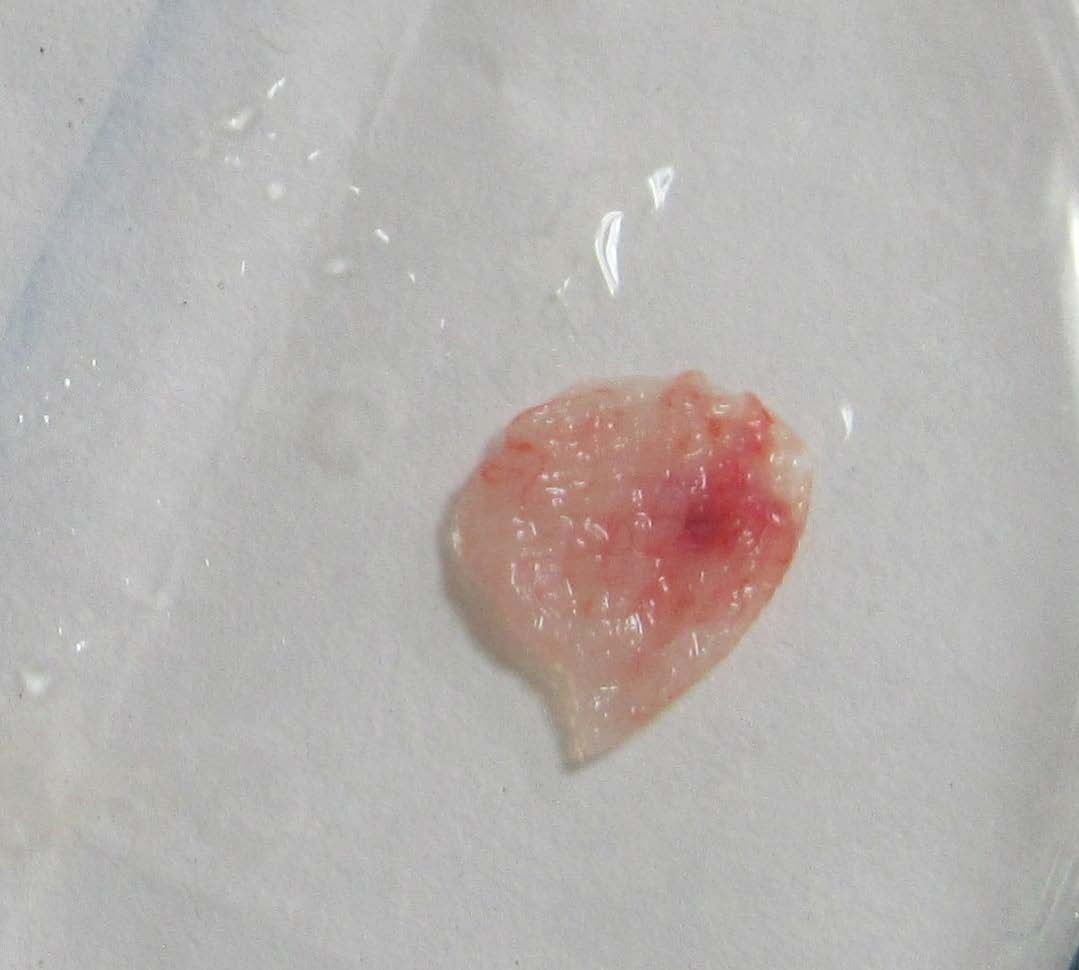
This letter presents simple novel technique during implant placement that ensures effective, precise placement and retention of Platelet Rich Fibrin (PRF) membrane at juxta-crestal bone around dental implant in the maxillary aesthetic zone. During the surgical phase, fresh PRF preparation was done following standard protocol. Antecubital vein blood was collected in 10 ml test-tube without any anticoagulant and immediately subjected to centrifugation at 3000 rpm for 10-12 min. The procedure resulted in the formation of three layers namely; acellular plasma at topmost layer, fibrin clot in the middle of tube and red corpuscles at the bottom. The upper straw coloured layer was discarded and the middle fraction of fibrin clot collected and squeezed between gauze-piece to obtain PRF membrane [Table/Fig-1].
Freshly prepared PRF membrane.
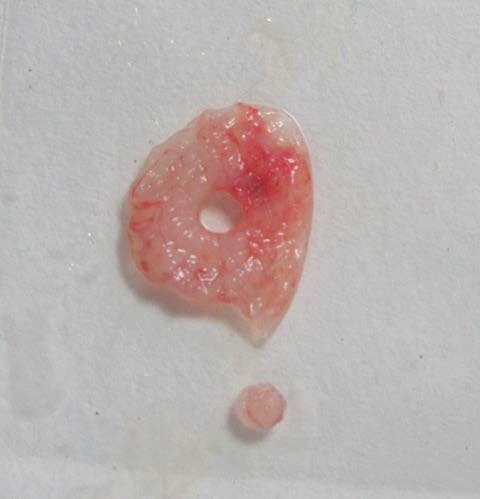
PRF membrane with a hole punched in the centre.
Customized PRF membrane placed around the collar of the dental implant in situ.

The prepared PRF membrane was positioned in the Petri dish and a tissue punch with diameter corresponding to that of the collar diameter of the dental implant was used to punch the membrane at the centre. This produced a PRF membrane with a hole in the centre that exactly matched with the crestal diameter of the dental implant used [Table/Fig-2].
The PRF membrane with hole was positioned around the dental implant inserted in the osteotomy site [Table/Fig-3]. It encircled the implant completely along the periphery and was snugly approximated at the crestal bone level.
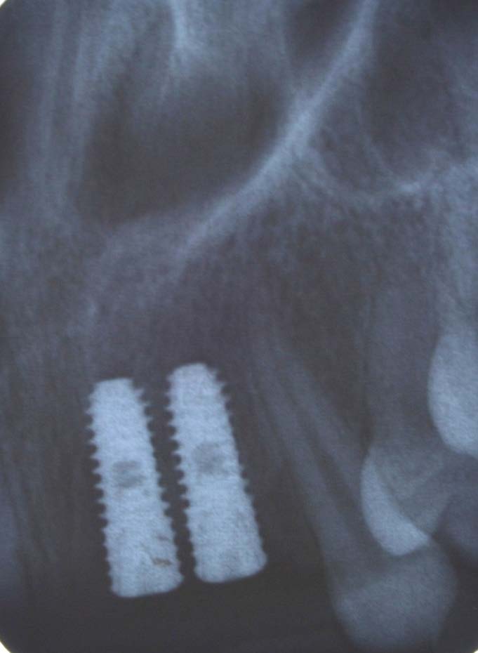
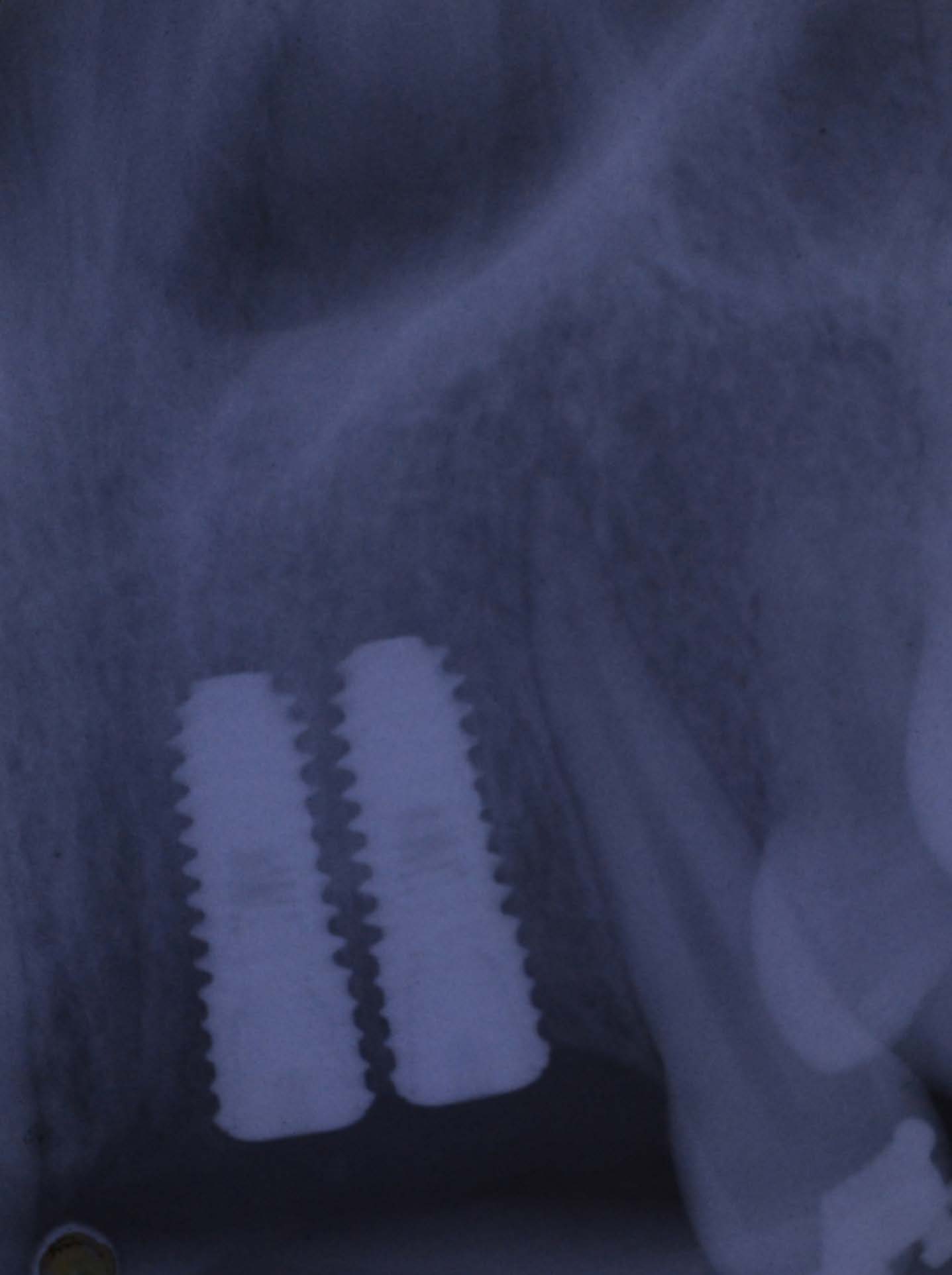
The complete encirclement around implant ensured non mobility of the PRF membrane and persistent positioning in close proximity to the crestal bone ensuring maximal beneficial effect in the crestal bone area during healing period [Table/Fig-4,5].
Intra oral peri apical radiograph depicting the crestal bone in early placed dental implant in post extraction site.
Intra oral peri apical radiograph depicting the crestal bone after three months of immediately placed dental implant augmented with Platelet Rich Fibrin (PRF) membrane at juxta-crestal bone around dental implant.
This simple clinical step during implant placement permits precise orientation and sustained positioning of the PRF membrane around implant delivering favourable healing factors around the most critical area around implant neck.
[1]. Ishikawa T, Kitajima H, Salama H, Salama M, Moroi H, Garber D, Three-dimensional bone and soft tissue requirements for optimizing esthetic results in compromised cases with multiple implantsInt J Periodontics Restorative Dent 2010 30:503-11. [Google Scholar]
[2]. Kiran NK, Mukunda KS, TilakRaj TN, Platelet concentrates:A promising innovation in DentistryJ Dent Sci Res 2011 2(1):50-61. [Google Scholar]
[3]. Kundu R, Rathee M, Effect of Platelet-Rich-Plasma (PRP) and implant surface topography on implant stability and boneJ Clin Diagn Res 2014 8(6):26-30. [Google Scholar]
[4]. Lee JW, Kim SG, Kim JY, Lee YC, Choi JY, Draqos R, Restoration of a peri-implant defect by platelet-rich fibrinOral Surg Oral Med Oral Pathol Oral Radiol Endod 2012 113(4):459-63. [Google Scholar]
[5]. Choukroun J, Diss A, Simonpieri A, Girard MO, Schoeffler C, Dohan SL, Platelet rich fibrin (PRF):A second-generation platelet concentrate. Part IV:clinical effects on tissue healingOral Surg Oral Med Oral Pathol Oral Radiol Endod 2006 101(3):56-60. [Google Scholar]
[6]. Boora P, Rathee M, Bhoria M, Effect of Platelet Rich Fibrin (PRF) on peri-implant soft tissue and crestal bone in one-stage implant placement:A randomized controlled trialJ Clin Diagn Res 2015 9(4):18-21. [Google Scholar]