Tracheal Agenesis with Tracheo-oesophageal Fistula
Somashekhar Marutirao Nimbalkar1, Vijay K Patel2, Dipen Vasudev Patel3, Ankur Rajinder Sethi4
1Professor, Department of Paediatrics,Pramukhswami Medical College, Karamsad, Gujarat, India.
2Senior Resident, Department of Paediatrics,Pramukhswami Medical College, Karamsad, Gujarat, India.
3Assistant Professor, Department of Paediatrics,Pramukhswami Medical College, Karamsad, Gujarat, India.
4Assistant Professor, Department of Paediatrics,Pramukhswami Medical College, Karamsad, Gujarat, India.
NAME, ADDRESS, E-MAIL ID OF THE CORRESPONDING AUTHOR: Dr. Somashekhar Nimbalkar, Professor, Department of Pediatrics, Shree Krishna Hospital, Pramukhswami Medical College, Karamsad, District: Anand, Gujarat-388325, India.
Phone: +919825087842,
E-mail:somu_somu@yahoo.com
Tracheal Agenesis (TA) presents with respiratory distress at birth. Diagnosis requires recognition of clinical signs in newborns like failure of endotracheal intubation, respiratory distress with absent air entry over both side of chest and inaudible cry. We describe a TA Floyd Type I with a Tracheo-Oesophageal Fistula (TOF) without other congenital malformations.
Tracheal agenesis, Tracheo-oesophageal fistula, Intubation, Respiratory distress
Introduction
Tracheal agenesis (TA) is a rare congenital defect with an unexpected emergency presentation. In 90% of presenting cases it exists with other congenital anomalies. Cardiovascular and gastrointestinal anomalies account for a significant percentage of these congenital anomalies. The incidence is approximately 1:50 000 with a male: female ratio of 2:1. TA may be a part of complex malformations with vertebral, anal, cardiovascular, tracheo-oesophageal, and renal and limb (VACTERL) anomalies [1-4]. There is association with prematurity and polyhydramnios [3]. The clinical symptoms are usually severe, and many cases die soon after birth. Floyd classified TA in three subtypes [5]. We describe a case of Floyd Type I with a distal TOF in a female newborn. Our objective to report this case is to provide information that can help the practitioner in case of difficulty in doing intubation.
Case Report
Human Research Ethics Committee of Shree Krishna Hospital, Karamsad, India approved this case for presentation. Informed consent was sought and the parents gave consent to the postmortem and subsequent publication. A 1,500 gm preterm (32 weeks maturity) female born by caesarean section to 30-year-old fourth gravida mother with no living child who had a pregnancy complicated by polyhydramnios. At birth there was gasping respiration and poor muscle tone. Heart rate was 100 after initial steps of resuscitation so baby required positive pressure ventilation (PPV) by bag and mask. Apgar score was 3 and 5 at 1 and 5 minutes respectively. On performing endotracheal intubation, larynx was visualized clearly up to vocal cord but 3 mm diameter endotracheal tube did not pass beyond the glottis on repeated attempts. So intubation with 2.5mm tube was attempted unsuccessfully. This too failed to pass beyond glottis, so PPV by bag and mask ventilation with 100% oxygen was continued with minimum chest expansion and poor air entry over both sides of chest with slight improvement in heart rate and colour. We passed feeding tube in to the stomach without encountering any difficulty. We considered possibilities of congenital laryngeal stenosis, congenital subglottic laryngeal web or TA.
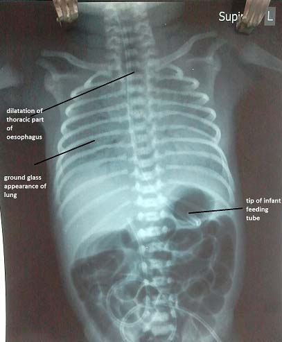
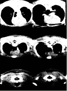
X-ray chest [Table/Fig-1] showed bilateral ground glass appearance with air-bronchogram. CT SCAN [Table/Fig-2] of neck was done which showed airway lumen up to C5 vertebral body and it appeared small in caliber ( at C4 level) with slit like laryngeal air way, this suggested sub-glottic air way stenosis. Lower part of tracheal lumen was visualized at D2 vertebral level which could be traced up to tracheal bifurcation and was associated with marked dilatation of thoracic part of oesophagus. There was also non-visualization of lower laryngeal air way lumen and trachea between C5 and D1 vertebral body.
During preparation for thoracic surgery baby died at the age of 6 hours. Histopathologic post-mortem showed obstructed laryngeal end due to anomalous cricoid cartilage. The part of trachea was absent with short distal trachea communicating to oesophagus via tracheo-oesophageal fistula. The oesophagus showed an elevated opening on the anterior wall which was communicating with the trachea. There were no external anomalies found. No other anomalies were seen in other organs during post mortem examination. So this was a case of abnormal cricoids cartilage with absent upper segment of trachea TA and a distal tracheo-oesophageal fistula.
X-Ray chest and abdomen shows ground glass appearance of lung
In the upper section of the chest at C7-D1 level (lower third of figure) there is no trachea seen but it is seen lower down at D2 level (middle third of figure) and the tracheal bifurcation is seen at D3 level
Discussion
In 1900 Payne described the first case of TA. Floyd et al described three different anatomic patterns of TA. Type I is characterized by absence of proximal trachea and by the presence of a distal tracheo-oesophageal fistula, type II is complete absence of trachea and presence of normal bifurcating bronchi. In type III, two main bronchi arise independently from the oesophagus [5]. The relative frequency of these types I, II, and III is 13%, 62%, and 25% respectively [6]. TA should be suspected in newborn with history of polyhydramnios, respiratory distress, absence of audible cry at birth and failure to intubate beyond the vocal cord [7]. The presenting symptoms and sign in our case were similar to other previously reported cases but there were no other anomalies in our case which were reported by many of them. The majority of the cases died at birth or shortly thereafter. Most cases do not survive long term because of associated other congenital malformation and failure to manage airway. The surgical management of neonates with TA is difficult. A systematic surgical approach does not exist but limited success of surgical management is reported and despite progress of surgical developments, the prognosis is poor [8,9]. Limited reports are available on long term survival in patient with TA. Few cases reported with long term survival where oesophagus was used as substitute for trachea [10,11]. TA without TOF can be diagnosed prenatally based on congenital high airway obstruction syndrome (CHAOS) on Ultrasonography and Prenatal MRI may provide a definitive diagnosis [12].
Conclusion
TA is a rare congenital anomaly which should be considered when the baby was born without crying, and intubation of a newborn baby is difficult. CT imaging is useful in diagnosis. Surgical management should be offered but even in skilled centres the outcomes may not be favourable.
[1]. JM Milstein, M Lau, RG Bickers, Tracheal agenesis in infants with VATER associationAm J Dis Child 1985 139:77-80. [Google Scholar]
[2]. JA Evans, CR Greenberg, L Erdile, Tracheal agenesis revisited: analysis of associated anomaliesAm J Med Genet 1999 82:415-22. [Google Scholar]
[3]. MB Van Veenendaal, KD Liem, HA Marres, Congenital absence of the tracheaEur J Pediatr 2000 159:8-13. [Google Scholar]
[4]. SR Mandrekar, S Amoncar, RG Pinto, Tracheal agenesis with broncho-esophageal fistula in VACTERL / TACRD associationIndian J of Hum Genet 2013 19(1):87-89. [Google Scholar]
[5]. S Ergun, T Tewfik, S Daniel, Tracheal agenesis: A rare but fatal congenital anomalyMcgill J Med 2011 13(1):10 [Google Scholar]
[6]. J Kerschner, DW Klotch, Tracheal agenesis:a case report and review of literatureOtolaryngol Head Neck Surg 1989 116:123-28. [Google Scholar]
[7]. B De Jose Maria, R Drudis, E Monclus, A Silva, S Santander, V Cusí, Management of tracheal agenesisPaediatric Anaesthesia 2000 10:441-44. [Google Scholar]
[8]. E Hiyama, T Yokoyama, T Ichikawa, Y Matsuura, Surgical management of tracheal agenesisJ Thorac Cardiovasc Surg. 1994 108:830-33. [Google Scholar]
[9]. N Usui, M Kamiyama, G Tani, Y Takama, H Soh, S Uehara, Three-stage reconstruction of the airway and alimentary tract in a case of tracheal agenesisAnn Thorac Surg 2010 89:2019-22. [Google Scholar]
[10]. Y Fuchimoto, M Mori, F Takasato, H Tomita, Y Yamamoto, N Shimojima, long-term survival case of tracheal agenesis: management for tracheoesophageal fistula and esophageal reconstructionPediatr Surg Int 2011 27:103-06. [Google Scholar]
[11]. H Soh, H Kawahawa, K Imura, M Yagi, A Yoneda, A Kubota, Tracheal agenesis in a child who survived for 6 yearsJ Pediatr Surg 1999 34(10):1541-43. [Google Scholar]
[12]. MD de Groot-van der Mooren, MC Haak, P Lakeman, TE Cohen-Overbeek, JP van der Voorn, JH Bretschneider, RM van Elburg, Tracheal agenesis: approach toward this severe diagnosis. Case report and review of the literatureEur J Pediatr. 2012 171(3):425-31. [Google Scholar]