A Rare Case of an Epidermoid Cyst in the Parotid Gland – which was Diagnosed by Fine Needle Aspiration Cytology
Panna N. Hegde1, Kishan Prasad H.L.2, Sunil Kumar Y.3, K. Sajitha4, Pooja Sarda Roy5, Mary Raju6, Vikram Shetty7
1 Post Graduate, Department of Pathology, K S Hegde Medical Academy, Mangalore, Karnataka, India.
2 Associate Professor, Department of Pathology, K S Hegde Medical Academy, Mangalore, Karnataka, India.
3 Associate Professor, Department of Pathology, K S Hegde Medical Academy, Mangalore, Karnataka, India.
4 Post Graduate, Dapartment of Pathology, K S Hegde Medical Academy, Mangalore, Karnataka, India.
5 Post Graduate, Department of Pathology, K S Hegde Medical Academy, Mangalore, Karnataka, India.
6 Post Graduate, Department of Pathology, K S Hegde Medical Academy, Mangalore, Karnataka, India.
7 Director, Nitte Meenakshi Institute of Cranio Facial Surgery, Mangalore, Karnataka, India.
NAME, ADRESS, E-MAIL ID OF THE CORESPONDING AUTHOR: Dr. Kishan Prasad, Associate Professor, Department of Pathology, K S Hegde Medical Academy of Nitte University, Deralakatte, Mangalore, Karnataka, India.
Phone: 09480503190,
E-mail: dr_kishanpath@yahoo.com.
Cystic lesions are common in the head and neck. The most common are the cutaneous cysts, which are referred to as epidermal cysts. These cysts present as nodular and fluctuant subcutaneous lesions and they are seen most commonly in the acne - prone areas like the head, neck and the back. They arise following a localized inflammation of the hair follicle and occasionally after the implantation of the epithelium, following a trauma or surgery. The presence of benign cystic lesions in the salivary glands is rare.We are presenting a rare case of a 55-year-old male who presented with a soft swelling on the left side of the face. A diagnosis of an epidermoid cyst was given on cytology. A superficial parotidectomy was performed and the histopathology confirmed the above diagnosis
Epidermal cyst, Epidermoid cyst, Salivary glands, FNAC
INTRODUCTION
Cystic lesions are uncommon within the parotid gland, which comprise approximately 5% of all the salivary gland tumours, a majority of which represent the cystic components of neoplasms [1]. An epidermoid cyst is a benign cyst which is most commonly seen in the skin. The cyst develops out of the ectodermal tissue. Histologically, it is made of a thin layer of squamous epithelium. Several synonyms exist for epidermoid cysts: epidermal cysts, epidermal inclusion cysts, infundibular cysts and keratin cysts. The epidermal inclusion cysts more specifically refer to the implantation of epidermal elements into the dermis. The infundibular cysts originate from the infundibular portion of the hair follicle. The presence of epidermoid cysts in the salivary gland is a rare entity and these cysts require surgical interventions. Hence, it is very essential to have a pre-operative diagnosis for the workup of the patients. In this case, the diagnosis of an epidermoid cyst was given on cytology and later, it was histopathologically confirmed. There are only very few case reports on epidermoid cysts which had occurred in parotid glands, which have been published in the world literature.
CASE HISTORY
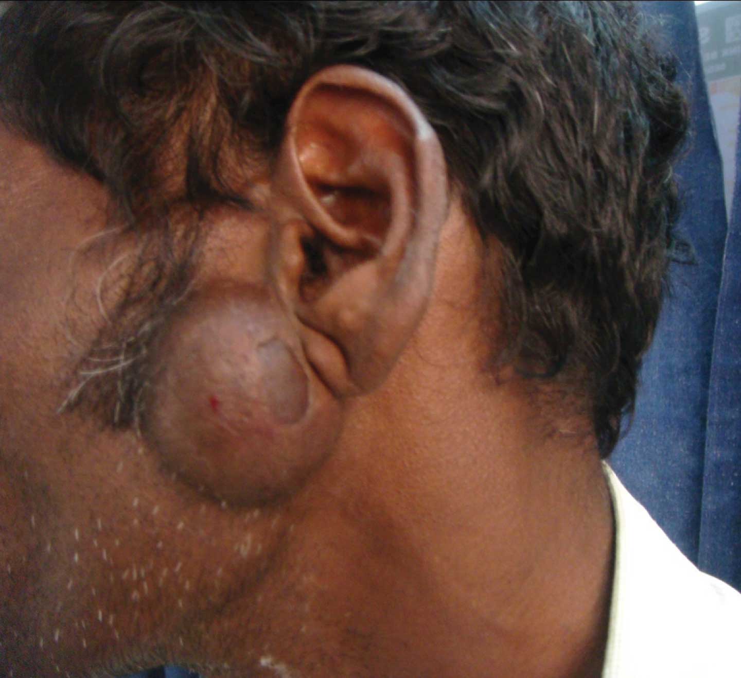
A 55-year-old male presented with a salivary gland swelling on the left side of the face, which was of two and a half year’s duration. The swelling was gradually increasing in size. His past history revealed a surgery which was done for the same, two and a half years ago, after which he had noticed an increase in the size of the swelling. On examination, a 4 x 3 cm, ovoid, mobile swelling was seen in the pre – auricular region. The skin over the swelling was scarred [Table/Fig-1]. The swelling was soft in consistency. An ultrasound examination showed an evidence of a hypoechoic cystic lesion in the left parotid region, which measured 3.1x1.3 cm. There was no vascularity in the lesion. The clinical impression was given as a lipoma over the left pre-auricular region.
Clinical photo of the salivary gland swelling
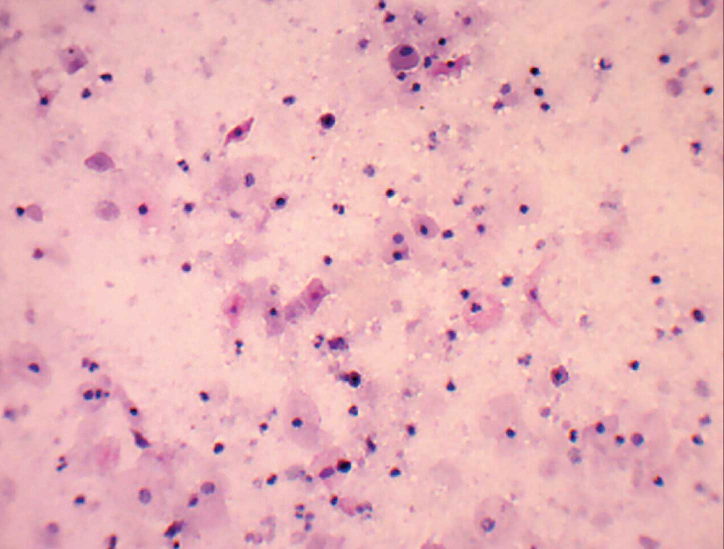
Fine needle aspiration of the swelling yielded a foul smelling, thick, pultaceous material. Its smears showed sheets of benign superficial and intermediate squamous cells in a background of a mild inflammatory infiltrate [Table/Fig-2]. A cytological diagnosis of an epidermoid cyst of the left parotid gland was made.
Cytology showing mature squamous cells and anucleatesquames. (MGG stain X100)
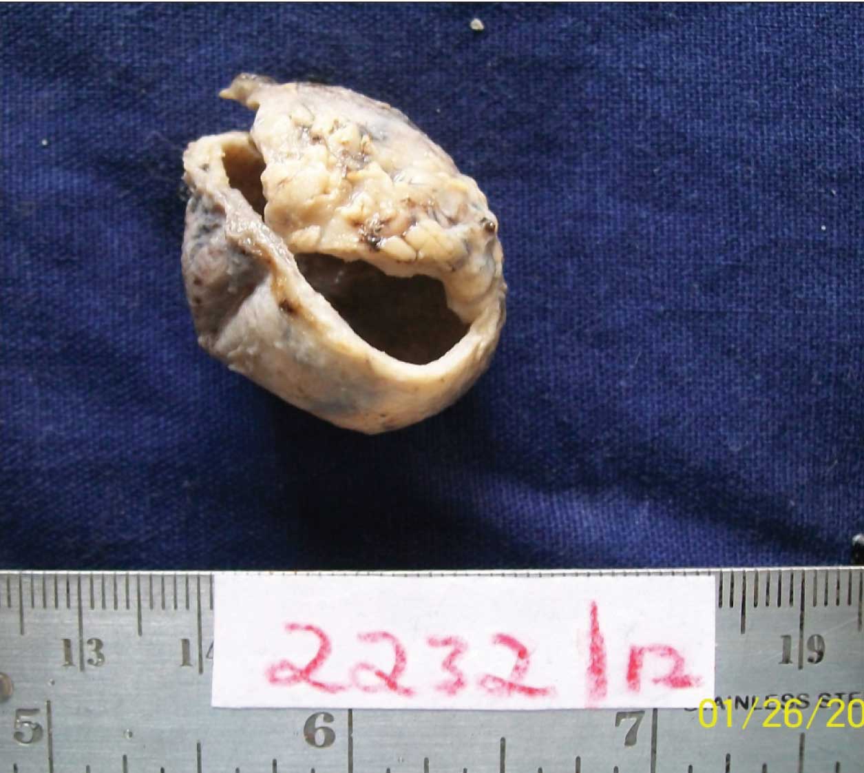
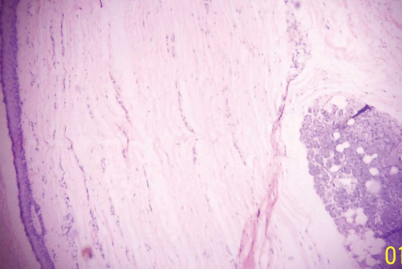
The patient underwent a superficial parotidectomy and the postoperative period was uneventful. The gross examination of the specimen showed a grey brown, globular mass which measured 3x2.5cm [Table/Fig-3]. Its cut surface yielded a pultaceous material. Sections were given from relevant areas in the cyst wall. Microscopy showed normal parotid acini with the adjacent cyst wall, which were lined by stratified squamous epithelium with an intarluminal laminated keratinous material [Table/Fig-4]. A final diagnosis of an epidermoid cyst of the left parotid gland was made. The patient is on regular follow up and is doing fine.
Gross picture showing superficial parotidectomy specimen with a cystic lesion
Histology showing parotid acini with adjacent cyst wall lined by squamous epithelium (H&E,X100).
DISCUSSION
Epidermoid cysts are common skin lesions that consist of epithelial lined cavities which are filled with viscous or semi solid epithelial degradation products. They are developmental cysts that occur in the head and neck with an incidence which ranges from 1.6 to 6.9% [2,3]. Epidermoid cysts usually occur secondary to obstructions, whereas dermoid cysts arise from developmental epithelial remnants or they are secondary to traumatic implantations of epithelial fragments [1].
The benign parotid cystic salivary lesions have a relatively low incidence [4].These cysts account for 5% of all the parotid lesions. Both men and women are equally affected and they are commonly seen between the fifth and seventh decades of life [1]. Clinically, they present as painless swellings without any attachment to the overlying skin or involvement of the facial nerve. The cystic lesions of the parotid can be either congenital or acquired. The congenital lesions are most often ectodermal in origin and they include branchial cleft cysts/lymphoepithelial cysts. The acquired cysts can be due to obstructions, neoplasms, calculi and trauma. The neoplasms include benign mixed lesions, Warthin’s tumour, mucoepidermoid carcinoma, adenoid cystic carcinoma and andacinic cell carcinoma, all of which can present as cystic lesions [5].
An epidermoid cyst of the parotid gland is a rare benign cystic lesion that requires a surgical intervention. It is derived from the epidermis and is formed by a cystic enclosure of the epithelium within the dermis, that becomes filled with keratin and lipid – rich debris [2]. It is common in young to middle aged adults.It can occur at the site of surgical incisions due to an iatrogenic implantation of the epidermis into the deeper tissues [4,5]. Its clinical and radiological characteristics can be ambiguous. Its histology shows predominantly squamous cells. Such lesions are quite unusual and they are not included in the WHO classification [4].
The diagnosis of cystic lesions is challenging, owing to the difficulty of determining the benign versus the malignant processes. Malignant lesions are frequently suspected when there is a rapid enlargement which is associated lymphadenopathy or facial nerve paresis. This distinction plays an important role in determining the treatment modality. The preoperative diagnosis of the lesion plays an important role. Fine needle aspiration cytology is the most reliable and the least expensive method which helps in making a preoperative diagnosis.
CONCLUSION
To conclude, epidermoid cysts of the parotid gland are rare entities, with only few cases having been mentioned in the literature. Epidermoid cysts can be considered as a differential diagnosis in cases with a recurrent, painless enlargement of the parotid gland which has a soft consistency. Our case has explained the significance of an accurate pre-operative diagnosis of a benign cystic lesion of the parotid and its significance in its further management.
[1]. Baschinsky D, Hameed A, Kehyani S, Fine needle aspiration cytological features of Dermoid cyst of parotid gland: A report of two casesDiagnostic cytopathology 1999 20(6):387-88. [Google Scholar]
[2]. Turki IM, Keddour AK, Daniel G, Dermoid cyst of the submandibular spaceFr ORL 2005 89:167-69. [Google Scholar]
[3]. Sanglee C, Kaun Kin H, Iatrogenic epidermoid cyst in the parotid gland-A case reportJ Korean Association Oral Maxillofacial Surg 2011 37:237-40. [Google Scholar]
[4]. Nagao T, Serizawa H, Iwaya K, Keratocystoma of the parotid gland: A report of two cases of an unusual pathologic entityMod Pathol 2001 15(9):1005-10. [Google Scholar]
[5]. Perkins M, Review of Fine needle aspiration cytology of salivary gland neoplasms with emphasis on differential diagnosisAm J Clin Pathol 2002 118:100-15. [Google Scholar]